| Midbrain tegmentum | |
|---|---|
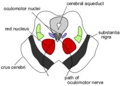
 Transverse section of mid-brain at level of superior colliculi. ("Tegmentum" visible center right.) | |
 Section through superior colliculus showing path of oculomotor nerve. (Tegmentum not labeled, but surrounding structures more clearly defined.) | |
| Details | |
| Part of | Midbrain |
| Identifiers | |
| Latin | Tegmentum Mesencephali |
| Anatomical terms of neuroanatomy | |
The midbrain is also known as the Mesencephalon and is one of the three major brain divisions. The midbrain is broken up into the tectum and the tegmentum. The midbrain tegmentum is the part of the midbrain extending from the substantia nigra to the cerebral aqueduct in a horizontal section of the midbrain. It forms the floor of the midbrain that surrounds the cerebral aqueduct. There are some really important structures located within the tegmentum. Additional structures include the reticular formation, red nucleus, ventral tegmental area (VTA) and the periaqueductal grey matter. The midbrain tegmentum contains thousands of neurons that are responsible for a variety of many functions, two of which include the control of movement and sensory systems. [1]
The Drug and Reward Pathway edit
Researchers have found that the ventral tegmental area (VTA), which is a part of the midbrain tegmentum, has played a key role in addiction and reward [2]. One important component of the drug-reward circuitry is the glutamate neuronal system. This system directly effects the dopamine system as well. Moreover, it has been found that when it comes to addiction specifically, it is the Glutaminergic input from the prefrontal cortex, hippocampal formation and the basolateral amygdala that are responsible. These input signals end up in the ventral tegmental area as well as the nucleus accumbens (NAcc)[2]. The VTA has been associated with another pathway known as the mesocorticolimbic DA (dopamine) pathway. The VTA is a region of the brain involved in locomotor functions and drug self-administration through the projection of the VTA to the nucleus accumbens (NAcc)[3].
Sexual Behaviour edit
The midbrain tegmentum will receive input from a region of the brain known as the medial preoptic area. One study conducted research on lesions to this area to determine what functions may be affected as a result. One thing that the researchers found was that when they induced lesions in the bilateral medial preoptic regions, it inhibited male sexual behavior [4]. They also found that when they directly produced a lesion in the dorsolateral tegmentum, a region in the midbrain tegmentum, it also inhibited male sexual behavior [4]. Therefore, these results suggest that any neural factors involved in male sexual behaviour either originate or pass through the tegmentum.
Diseases and Disorders edit
One autosomal recessive disease known to be correlated with the midbrain tegmentum is known as Wilson’s disease. This is a rare disease which effects your metabolism by accumulating excessive amounts of copper in the liver, brain and other tissues [5]. The midbrain tegmentum of a patient suffering from Wilson’s disease will occasionally be affected. A biomarker for this is the “double panda sign” which will present itself on the midbrain tegmentum on an MRI scan [6].
Several researchers have found a correlation of the midbrain tegmentum with a variety of headaches. More specifically, it has been found that the activation of cluster headaches (CH) occurs in the midbrain tegmentum [7]. This may be occurring because the midbrain tegmentum, in conjunction with the periaqueductal gray area, is involved in the signaling processes of pain. Moreover, using PET scans, it has been found that two other headaches have been associated with the midbrain tegmentum. Specifically, the activations of Hemicrania continua and Paroxysmal hemicrania, two rare forms of headaches, occur mainly in the posterior hypothalamic region of the brain [7]. However the activation of these two rare forms of headaches extend posteriorly into the midbrain tegmentum [7].
See also edit
External links edit
- Photo
- "Anatomy diagram: 13048.000-3". Roche Lexicon - illustrated navigator. Elsevier. Archived from the original on 2014-01-01.
Notes edit
- ^ Paradiso, Mark F. Bear ; Barry W. Connors ; Michael A. (2012). Neuroscience : exploring the brain (4. ed. ed.). Philadelphia [u.a.]: Lippincott Williams & Wilkins. p. 200. ISBN 978-0-7817-7817-6.
{{cite book}}:|edition=has extra text (help)CS1 maint: multiple names: authors list (link) - ^ a b MORGANE, P; GALLER, J; MOKLER, D (February 2005). "A review of systems and networks of the limbic forebrain/limbic midbrain". Progress in Neurobiology. 75 (2): 143–160. doi:10.1016/j.pneurobio.2005.01.001.
- ^ Vezina, Paul (January 2004). "Sensitization of midbrain dopamine neuron reactivity and the self-administration of psychomotor stimulant drugs". Neuroscience & Biobehavioral Reviews. 27 (8): 827–839. doi:10.1016/j.neubiorev.2003.11.001.
- ^ a b Brackett, Nancy L.; Edwards, David A. (January 1984). "Medial preoptic connections with the midbrain tegmentum are essential for male sexual behavior". Physiology & Behavior. 32 (1): 79–84. doi:10.1016/0031-9384(84)90074-X.
- ^ Rodriguez-Castro, Kryssia Isabel (2015). "Wilson's disease: A review of what we have learned". World Journal of Hepatology. 7 (29): 2859. doi:10.4254/wjh.v7.i29.2859.
{{cite journal}}: CS1 maint: unflagged free DOI (link) - ^ Kakkar, Chandan; Kakkar, Shruti; Saggar, Kavita; Goraya, Jatinder S.; Ahluwalia, Archana; Arora, Ankur (23 May 2016). "Paediatric brainstem: A comprehensive review of pathologies on MR imaging". Insights into Imaging. 7 (4): 505–522. doi:10.1007/s13244-016-0496-3.
- ^ a b c Matharu, Manjit S.; Zrinzo, Ludvic (24 February 2010). "Deep Brain Stimulation in Cluster Headache: Hypothalamus or Midbrain Tegmentum?". Current Pain and Headache Reports. 14 (2): 151–159. doi:10.1007/s11916-010-0099-5.
This is a user sandbox of Marcmitri. You can use it for testing or practicing edits. This is not the sandbox where you should draft your assigned article for a dashboard.wikiedu.org course. To find the right sandbox for your assignment, visit your Dashboard course page and follow the Sandbox Draft link for your assigned article in the My Articles section. |