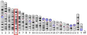
Homer protein homolog 1 or Homer1 is a neuronal protein that in humans is encoded by the HOMER1 gene.[5][6][7] Other names are Vesl and PSD-Zip45.
Structure
editHomer1 protein has an N-terminal EVH1 domain, involved in protein interaction, and a C-terminal coiled-coil domain involved in self association. It consists of two major splice variants, short-form (Homer1a) and long-form (Homer1b and c). Homer1a has only EVH1 domain and is monomeric while Homer1b and 1c have both EVH1 and coiled-coil domains and are tetrameric.[8][9] The coiled-coil can be further separated into N-terminal half and C-terminal half. The N-terminal half of the coiled-coil domain is predicted to be a parallel dimer while the C-terminus half is a hybrid of dimeric and anti-parallel tetrameric coiled-coil. As a whole, long Homer is predicted to have a dumbbell-like structure where two pairs of EVH1 domains are located on two sides of long (~50 nm) coiled-coil domain.[9] Mammals have Homer2 and Homer3, in addition to Homer1, which have similar domain structure. They also have similar alternatively spliced forms.
Tissue distribution
editHomer1 is expressed widely in the central nervous system as well as peripheral tissue including heart, kidney, ovary, testis, and skeletal muscle. Subcellularly in neurons, Homer1 is concentrated in postsynaptic structures and constitutes a major part of the postsynaptic density.
Function
editEVH1 domain interacts with PPXXF motif. This sequence motif exists in group 1 metabotrophic glutamate receptor (mGluR1 and mGluR5), IP3 receptors (IP3R), Shank, transient receptor potential canonical (TRPC) family channels, drebrin, oligophrenin, dynamin3, CENTG1, and ryanodin receptor.[5][7][10][11][12][13] Through its tetrameric structure, long forms of Homer (such as Homer1b and Homer1c) are proposed to cross link different proteins. For example, group 1 mGluR is crossed linked with its signaling downstream, IP3 receptor.[10] Also, through crosslinking another multimeric protein Shank, it is proposed to comprise a core of the postsynaptic density.[9]
Notably, the expression of Homer1a is induced by neuronal activity while that of Homer1b and 1c are constitutive. Thus Homer1a is classified as an immediate early gene. Homer1a, acts as a natural dominant negative form that blocks interaction between long-forms and their ligand proteins by competing with the EVH1 binding site on the ligand proteins. In this way, the short form of Homer uncouples mGluR signaling and also shrinks dendritic spine structure.[6][14] Therefore, the short form of Homer is considered to be a part of a mechanism of homeostatic plasticity that dampens the neuronal responsiveness when input activity is too high. The long form Homer1c plays a role in synaptic plasticity and the stabilization of synaptic changes during long-term potentiation.[15]
The coiled-coil domain is reported to interact with syntaxin13 and activated Cdc42. The interaction with Cdc42 inhibit the activity of Cdc42 to remodel dendritic spine structure.
Rapid antidepressant effects
editHomer1a switches mGluR5 signaling to increase AMPA receptor activity for the rapid antidepressant actions of sleep deprivation.[16]
See also
editReferences
edit- ^ a b c GRCh38: Ensembl release 89: ENSG00000152413 – Ensembl, May 2017
- ^ a b c GRCm38: Ensembl release 89: ENSMUSG00000007617 – Ensembl, May 2017
- ^ "Human PubMed Reference:". National Center for Biotechnology Information, U.S. National Library of Medicine.
- ^ "Mouse PubMed Reference:". National Center for Biotechnology Information, U.S. National Library of Medicine.
- ^ a b Shiraishi-Yamaguchi Y, Furuichi T (2007). "The Homer family proteins". Genome Biology. 8 (2): 206. doi:10.1186/gb-2007-8-2-206. PMC 1852408. PMID 17316461.
- ^ a b Tu JC, Xiao B, Yuan JP, Lanahan AA, Leoffert K, Li M, et al. (October 1998). "Homer binds a novel proline-rich motif and links group 1 metabotropic glutamate receptors with IP3 receptors". Neuron. 21 (4): 717–726. doi:10.1016/S0896-6273(00)80589-9. PMID 9808459. S2CID 2851554.
- ^ a b "Entrez Gene: HOMER1 homer homolog 1 (Drosophila)".
- ^ Hayashi MK, Ames HM, Hayashi Y (August 2006). "Tetrameric hub structure of postsynaptic scaffolding protein homer". The Journal of Neuroscience. 26 (33): 8492–8501. doi:10.1523/JNEUROSCI.2731-06.2006. PMC 6674353. PMID 16914674.
- ^ a b c Hayashi MK, Tang C, Verpelli C, Narayanan R, Stearns MH, Xu RM, et al. (April 2009). "The postsynaptic density proteins Homer and Shank form a polymeric network structure". Cell. 137 (1): 159–171. doi:10.1016/j.cell.2009.01.050. PMC 2680917. PMID 19345194.
- ^ a b Xiao B, Tu JC, Petralia RS, Yuan JP, Doan A, Breder CD, et al. (October 1998). "Homer regulates the association of group 1 metabotropic glutamate receptors with multivalent complexes of homer-related, synaptic proteins". Neuron. 21 (4): 707–716. doi:10.1016/S0896-6273(00)80588-7. PMID 9808458. S2CID 16431031.
- ^ Rong R, Ahn JY, Huang H, Nagata E, Kalman D, Kapp JA, et al. (November 2003). "PI3 kinase enhancer-Homer complex couples mGluRI to PI3 kinase, preventing neuronal apoptosis". Nature Neuroscience. 6 (11): 1153–1161. doi:10.1038/nn1134. PMID 14528310. S2CID 807407.
- ^ Hwang SY, Wei J, Westhoff JH, Duncan RS, Ozawa F, Volpe P, et al. (August 2003). "Differential functional interaction of two Vesl/Homer protein isoforms with ryanodine receptor type 1: a novel mechanism for control of intracellular calcium signaling". Cell Calcium. 34 (2): 177–184. doi:10.1016/S0143-4160(03)00082-4. PMID 12810060.
- ^ Feng W, Tu J, Yang T, Vernon PS, Allen PD, Worley PF, Pessah IN (November 2002). "Homer regulates gain of ryanodine receptor type 1 channel complex". The Journal of Biological Chemistry. 277 (47): 44722–44730. doi:10.1074/jbc.M207675200. PMID 12223488.
- ^ Sala C, Futai K, Yamamoto K, Worley PF, Hayashi Y, Sheng M (July 2003). "Inhibition of dendritic spine morphogenesis and synaptic transmission by activity-inducible protein Homer1a". The Journal of Neuroscience. 23 (15): 6327–6337. doi:10.1523/JNEUROSCI.23-15-06327.2003. PMC 6740555. PMID 12867517.
- ^ Meyer D, Bonhoeffer T, Scheuss V (April 2014). "Balance and stability of synaptic structures during synaptic plasticity". Neuron. 82 (2): 430–443. doi:10.1016/j.neuron.2014.02.031. PMID 24742464.
- ^ Albert PR (October 2019). "Targeting Homer1a for Rapid Antidepressant Effects". Neuron. 104 (2): 182–183. doi:10.1016/j.neuron.2019.10.003. PMID 31647890. S2CID 204837754.
Further reading
edit- Xiao B, Tu JC, Worley PF (June 2000). "Homer: a link between neural activity and glutamate receptor function". Current Opinion in Neurobiology. 10 (3): 370–374. doi:10.1016/S0959-4388(00)00087-8. PMID 10851183. S2CID 8699597.
- Maruyama K, Sugano S (January 1994). "Oligo-capping: a simple method to replace the cap structure of eukaryotic mRNAs with oligoribonucleotides". Gene. 138 (1–2): 171–174. doi:10.1016/0378-1119(94)90802-8. PMID 8125298.
- Hillier LD, Lennon G, Becker M, Bonaldo MF, Chiapelli B, Chissoe S, et al. (September 1996). "Generation and analysis of 280,000 human expressed sequence tags". Genome Research. 6 (9): 807–828. doi:10.1101/gr.6.9.807. PMID 8889549.
- Brakeman PR, Lanahan AA, O'Brien R, Roche K, Barnes CA, Huganir RL, Worley PF (March 1997). "Homer: a protein that selectively binds metabotropic glutamate receptors". Nature. 386 (6622): 284–288. Bibcode:1997Natur.386..284B. doi:10.1038/386284a0. PMID 9069287. S2CID 4346579.
- Suzuki Y, Yoshitomo-Nakagawa K, Maruyama K, Suyama A, Sugano S (October 1997). "Construction and characterization of a full length-enriched and a 5'-end-enriched cDNA library". Gene. 200 (1–2): 149–156. doi:10.1016/S0378-1119(97)00411-3. PMID 9373149.
- Tu JC, Xiao B, Naisbitt S, Yuan JP, Petralia RS, Brakeman P, et al. (July 1999). "Coupling of mGluR/Homer and PSD-95 complexes by the Shank family of postsynaptic density proteins". Neuron. 23 (3): 583–592. doi:10.1016/S0896-6273(00)80810-7. PMID 10433269. S2CID 16429070.
- Roche KW, Tu JC, Petralia RS, Xiao B, Wenthold RJ, Worley PF (September 1999). "Homer 1b regulates the trafficking of group I metabotropic glutamate receptors". The Journal of Biological Chemistry. 274 (36): 25953–25957. doi:10.1074/jbc.274.36.25953. PMID 10464340.
- Minakami R, Kato A, Sugiyama H (June 2000). "Interaction of Vesl-1L/Homer 1c with syntaxin 13". Biochemical and Biophysical Research Communications. 272 (2): 466–471. doi:10.1006/bbrc.2000.2777. PMID 10833436.
- Ango F, Prézeau L, Muller T, Tu JC, Xiao B, Worley PF, et al. (June 2001). "Agonist-independent activation of metabotropic glutamate receptors by the intracellular protein Homer". Nature. 411 (6840): 962–965. doi:10.1038/35082096. PMID 11418862. S2CID 4417727.
- Wistow G, Bernstein SL, Wyatt MK, Fariss RN, Behal A, Touchman JW, et al. (June 2002). "Expressed sequence tag analysis of human RPE/choroid for the NEIBank Project: over 6000 non-redundant transcripts, novel genes and splice variants". Molecular Vision. 8: 205–220. PMID 12107410.
- Feng W, Tu J, Yang T, Vernon PS, Allen PD, Worley PF, Pessah IN (November 2002). "Homer regulates gain of ryanodine receptor type 1 channel complex". The Journal of Biological Chemistry. 277 (47): 44722–44730. doi:10.1074/jbc.M207675200. PMID 12223488.
- Hwang SY, Wei J, Westhoff JH, Duncan RS, Ozawa F, Volpe P, et al. (August 2003). "Differential functional interaction of two Vesl/Homer protein isoforms with ryanodine receptor type 1: a novel mechanism for control of intracellular calcium signaling". Cell Calcium. 34 (2): 177–184. doi:10.1016/S0143-4160(03)00082-4. PMID 12810060.
- Norton N, Williams HJ, Williams NM, Spurlock G, Zammit S, Jones G, et al. (July 2003). "Mutation screening of the Homer gene family and association analysis in schizophrenia". American Journal of Medical Genetics. Part B, Neuropsychiatric Genetics. 120B (1): 18–21. doi:10.1002/ajmg.b.20032. PMID 12815733. S2CID 25053099.
- Westhoff JH, Hwang SY, Duncan RS, Ozawa F, Volpe P, Inokuchi K, Koulen P (September 2003). "Vesl/Homer proteins regulate ryanodine receptor type 2 function and intracellular calcium signaling". Cell Calcium. 34 (3): 261–269. doi:10.1016/S0143-4160(03)00112-X. PMID 12887973.
- Yuan JP, Kiselyov K, Shin DM, Chen J, Shcheynikov N, Kang SH, et al. (September 2003). "Homer binds TRPC family channels and is required for gating of TRPC1 by IP3 receptors". Cell. 114 (6): 777–789. doi:10.1016/S0092-8674(03)00716-5. PMID 14505576. S2CID 10552676.
- Rong R, Ahn JY, Huang H, Nagata E, Kalman D, Kapp JA, et al. (November 2003). "PI3 kinase enhancer-Homer complex couples mGluRI to PI3 kinase, preventing neuronal apoptosis". Nature Neuroscience. 6 (11): 1153–1161. doi:10.1038/nn1134. PMID 14528310. S2CID 807407.


