Wikipedia:WikiProject Palaeontology/Paleoart review/Archive 1

As a start up, here's a restoration of Osteodontornis which Dysmorodrepanis points out has a too short beak in the caption. Are there any other problems with it? Since Arthur Weasly, who created the image, has left Wikipedia, anyone else could fix it instead, due to the license it is released under. I could do it if someone could send me some references for the beak. FunkMonk (talk) 13:27, 19 August 2009 (UTC)
- Simple, use the photo here as a basis. This may not necessarily be Osteodontornis (it is almost certainly not O. orri), but the proportions are the same. The head of the large pseudotooth birds, as far as is known, was about 25% skull, 75% beak. According to Mayr (2008) "A skull of the giant bony-toothed bird Dasornis", etc, the head of O. orri is known from an almost complete specimen, which has a length of 40 cm including the beak. Comparing with the Dasornis skull specimen (which has the beak missing but is almost the same size as the O. orri specimen) gives the same 25%-75% proportions. The exact figures for O. orri should be in the species' description, but this is unfortunately not available online. You can still use Howard's description as a reference, but it is probably better to cite Mayr (which will probably come online as free fulltext sooner or later, and is available as abstract even now). Dysmorodrepanis (talk) 14:11, 19 August 2009 (UTC)
- Thanks, I'll fix it one of these days, and post it up for review again. By the way, is the beak on this restoration accurate? http://www.dinozaury.com/images/rsgallery/original/pelagornithidae1_2.jpg FunkMonk (talk) 14:31, 19 August 2009 (UTC)
- In size yes. Like here, some reconstructions put a "nail" at the tip, as in pelicans, and this seems to be correct in fact (and is phylogenetically intriguing). See also figure 4b here and here (hmmm do I spot intraorbital salt glands?) - you can take the figure from the paper and use it as an underlay in Photoshop or whatever you use, rotate and flip it so that it matches the drawing, to check for bill accuracy and position the nostril. I wouldn't make the bill patterned as they did in the drawing - if they were Galloanseres it might have been so (check out any goose), but even if they were there is no need for it to be so. The nondescript coloration is rather appropriate; nothing about it is likely to be utterly wrong.Dysmorodrepanis (talk) 20:55, 19 August 2009 (UTC)
- I put this one together in a few minutes, what do you think? http://img99.imageshack.us/img99/9195/44406947.jpg FunkMonk (talk) 19:26, 24 August 2009 (UTC)
- The "teeth" would be one small between each two large - the info in the artifcle is shaky at best; it's frm a 2nd rate source I did not see. True, there is more than one than one small "tooth" between each 2 large, but only one of them would be conspicuous at the image's resolution. The others would just be "needletip points" (or whatever Howard apparently called them).
- But that's all I can see from here. Dysmorodrepanis (talk) 01:12, 25 August 2009 (UTC)
- How about this? http://img48.imageshack.us/img48/4220/65656116.jpg FunkMonk (talk) 16:46, 27 August 2009 (UTC)
- One can always tweak this or that - for example, the iris was more likely to be dark brown, but that's sheer statistics, not evidence-based; yellow is entirely possible too (blue would have been highly unlikely for example). You might check out Osteodontornis#External_links - the photos of SBMNH 309 in particular - and see if you note something you think should be in there, but I can't really see any reason not to leave it as it is. Thanks for the quick work! Dysmorodrepanis (talk) 21:04, 28 August 2009 (UTC)
- How about this? http://img48.imageshack.us/img48/4220/65656116.jpg FunkMonk (talk) 16:46, 27 August 2009 (UTC)
- I put this one together in a few minutes, what do you think? http://img99.imageshack.us/img99/9195/44406947.jpg FunkMonk (talk) 19:26, 24 August 2009 (UTC)
- In size yes. Like here, some reconstructions put a "nail" at the tip, as in pelicans, and this seems to be correct in fact (and is phylogenetically intriguing). See also figure 4b here and here (hmmm do I spot intraorbital salt glands?) - you can take the figure from the paper and use it as an underlay in Photoshop or whatever you use, rotate and flip it so that it matches the drawing, to check for bill accuracy and position the nostril. I wouldn't make the bill patterned as they did in the drawing - if they were Galloanseres it might have been so (check out any goose), but even if they were there is no need for it to be so. The nondescript coloration is rather appropriate; nothing about it is likely to be utterly wrong.Dysmorodrepanis (talk) 20:55, 19 August 2009 (UTC)
- Thanks, I'll fix it one of these days, and post it up for review again. By the way, is the beak on this restoration accurate? http://www.dinozaury.com/images/rsgallery/original/pelagornithidae1_2.jpg FunkMonk (talk) 14:31, 19 August 2009 (UTC)
The same image is also being used in the Pelagornithidae article, and in the List of fossil birds article, where is also identified as a Osteodontornis. Nightscream (talk) 05:30, 6 December 2009 (UTC)


I have been updating this image to be better in both accuracy and aesthetics. But I want your harshest of criticisms about this image, if it is ugly, if it is inaccurate in some way, something. Please tell me if there is anything you don't like about this image and I will improve it. Thanks! Giant Blue Anteater (talk) 19:46, 19 August 2009 (UTC)
- I don't really know anything about fish, but could you post the references you used, also for Ichthyostega maybe? Then I could at least check shapes and proportions. FunkMonk (talk) 19:32, 24 August 2009 (UTC)
- Thanks for responding. This is the image that I used for reference. The top one is Myllokunmingia, and the bottom is Haikouichthys. Oh and I just forgot that I drew Myllokunmingia as well, so I'm posting that image here as well. Stupid me for forgetting! As for Ichthyostega, I used Ahlberg et al.'s skeletal reconstruction as a reference, which can be found here. Giant Blue Anteater (talk) 20:07, 24 August 2009 (UTC)
August 2009 (UTC)
- I based the scales on its ancestors being scaly. It's like feathering an Eoraptor based on its descendants having some integumentation. Perhaps the scales of Ichthyostega were soft and therefore didn't preserve. Giant Blue Anteater (talk) 21:00, 25 August 2009 (UTC)
- Oh, I meant more like what kind of scales did you base them on? FunkMonk (talk) 09:56, 29 August 2009 (UTC)
- Well... I was just carelessly drawing the scales, without regard to proper shape. I will redraw the scales as properly rhombus-shaped like a fish's. Giant Blue Anteater (talk) 18:43, 13 September 2009 (UTC)
- Oh, I meant more like what kind of scales did you base them on? FunkMonk (talk) 09:56, 29 August 2009 (UTC)
- I based the scales on its ancestors being scaly. It's like feathering an Eoraptor based on its descendants having some integumentation. Perhaps the scales of Ichthyostega were soft and therefore didn't preserve. Giant Blue Anteater (talk) 21:00, 25 August 2009 (UTC)

I have decided to make a new illustration of Ichthyostega, covered with scales rather than slippery skin since it was more like a fish with legs than a primitive salamander as many illustrations including Arthur Weasley's depict it as. It simply doesn't make any sense for sarcopterygians to lose their scales yet regain them once they start laying eggs on land. Although I am aware that there is no evidence for scales on Ichthyostega and kin, but that does not mean that they lacked them, since they do come from an ancestor with scales. Your thoughts? Giant Blue Anteater (talk) 07:09, 22 August 2009 (UTC)
- I'm no expert on early tetrapods, but I'm pretty sure that fish scales originate from the dermis, while reptilian scales are epidermal. Wouldn't that mean that the scales of amniotes were independantly evolved after the fish-type scales of dermal origin were lost? That's not to say that Ichthyostega did not have these fish-like scales: it is still possible that it was not until more derived forms than Ichthyostega appeared that early tetrapods lost these types of scales. However, I don't yet know of any publications that describe early tetrapod integumentary structure, so don't take my word for it. Smokeybjb (talk) 18:35, 25 August 2009 (UTC)
- By looking at this[1], and on fish scales in general, shouldn't the scales be more overlapping? Now they're more like reptile scales it seems. FunkMonk (talk) 20:27, 25 August 2009 (UTC)
- Well, this is my first attempt on drawing scales ever, so don't expect any sapient being to be perfect. I'll keep practicing though. Giant Blue Anteater (talk) 21:13, 25 August 2009 (UTC)
- Basal synapsids kept these epidermal scales but lost them as they evolved glandular skin, I think. Ophiacodon was perserved with dermal "belly scales" that are not homologous to the epidermal belly scales of squamates I believe. So why no basal tetrapods? Giant Blue Anteater (talk) 21:13, 25 August 2009 (UTC)
- I can't find much on integumentary evolution in early tetrapods at the moment, but I found an online article that seems to suggest that a corneous layer formed over the dermoscutes (dermal scales of sarcopterygians) during early tetrapod evolution. This would imply that reptilian-grade scales could form on organisms over time while they still obtained the fish-like scales of their ancestors, and thus scales were never lost in amniotes until glandular skin appeared independantly in lissamphibians and derived synapsids.
- I found the information in "Dermo-epidermal interactions in reptilian scales: Speculations on the evolution of scales, feathers, and hairs" Journal of Experimental Zoology 302B(4). Unfortunately I cannot provide a link to the article as it is not open access. Smokeybjb (talk) 00:55, 26 August 2009 (UTC)
- The source from where I got the information about Ophiacodon's scales was from Darren Naish's answer to a question about synapsid skin on Ask a Biologist, with his reference being Carroll, R. L. 1969. Problems of the origin of reptiles. _Biological Reviews_ 44, 393-432. So the sources we gathered strengthens the case for a scaly Ichthyostega. The only problem left though are the scales themselves. As I said, this is my first attempt at drawing scales, but FunkMonk suggested that the scales should be overlapping like a fish. But however, the illustration of Tiktaalik dosen't show the detail of the scales that well, but can this be excused, or should I redraw the scales? Giant Blue Anteater (talk) 21:14, 26 August 2009 (UTC)
- Many life restorations of early tetrapods and tetrapodomorphs seem to depict them as having fish-like scales that are much smaller than the ones seen in the photograph of the Ichthyostega model that FunkMonk brought up. For example, see this fossil of Eusthenopteron[2]. I would say that the epidermal scales found in amniotes such as reptiles, which are generally circular in shape, probably did not yet occur in Ichthyostega. That being said the scales in your restoration seem to be a little reptilian in appearance. However, I'd think that the sort of mucus membrane found on fish, and presumably Ichthyostega, may have obscured these scales a bit. It's up to you whether you want to completely redraw the scales as overlapping. In my opinion, though, it would be easier to make the scales a little less prominent as to not have them look obviously reptilian. That way you wouldn't have to go through all that hard work replacing every one! Smokeybjb (talk) 23:36, 26 August 2009 (UTC)
- Yeah, putting some sort of layer over the skin that would blur the details out a bit could help with both making the individual scales less prominent, as well as making the surface look smoother, and more like a fish, so nothing would have to be redrawn. Here's a crude example I made in Photoshop, by simply marking part of the drawing that had scales and putting the "blur more" filter on: [3] FunkMonk (talk) 01:24, 27 August 2009 (UTC)
- Check out Tristichopteridae, their scales should be closest to the ones the ancestors of Ichthyostega inherited. Those of present-day coelacanths and even the Australian lungfish differ already - not that much different compared to a carp's for example, but still a bit. They are larger in the extant taxa too, but if we can exclude something in Ichthyostega then it's larger scales than those of Tristichopteridae ;-) In any case, they were Scale_(zoology)#Cosmoid_scales and thus unlike those of other fishes. Dysmorodrepanis (talk) 21:17, 28 August 2009 (UTC)
- Even though a filter for blurring things is available in Flash (the program of which I made this image in), I will redraw the scales as diamond-shaped like a fish's. In short, the image can wait. Giant Blue Anteater (talk) 20:09, 7 September 2009 (UTC)
- Yeah, putting some sort of layer over the skin that would blur the details out a bit could help with both making the individual scales less prominent, as well as making the surface look smoother, and more like a fish, so nothing would have to be redrawn. Here's a crude example I made in Photoshop, by simply marking part of the drawing that had scales and putting the "blur more" filter on: [3] FunkMonk (talk) 01:24, 27 August 2009 (UTC)
- Many life restorations of early tetrapods and tetrapodomorphs seem to depict them as having fish-like scales that are much smaller than the ones seen in the photograph of the Ichthyostega model that FunkMonk brought up. For example, see this fossil of Eusthenopteron[2]. I would say that the epidermal scales found in amniotes such as reptiles, which are generally circular in shape, probably did not yet occur in Ichthyostega. That being said the scales in your restoration seem to be a little reptilian in appearance. However, I'd think that the sort of mucus membrane found on fish, and presumably Ichthyostega, may have obscured these scales a bit. It's up to you whether you want to completely redraw the scales as overlapping. In my opinion, though, it would be easier to make the scales a little less prominent as to not have them look obviously reptilian. That way you wouldn't have to go through all that hard work replacing every one! Smokeybjb (talk) 23:36, 26 August 2009 (UTC)
- The source from where I got the information about Ophiacodon's scales was from Darren Naish's answer to a question about synapsid skin on Ask a Biologist, with his reference being Carroll, R. L. 1969. Problems of the origin of reptiles. _Biological Reviews_ 44, 393-432. So the sources we gathered strengthens the case for a scaly Ichthyostega. The only problem left though are the scales themselves. As I said, this is my first attempt at drawing scales, but FunkMonk suggested that the scales should be overlapping like a fish. But however, the illustration of Tiktaalik dosen't show the detail of the scales that well, but can this be excused, or should I redraw the scales? Giant Blue Anteater (talk) 21:14, 26 August 2009 (UTC)
- By looking at this[1], and on fish scales in general, shouldn't the scales be more overlapping? Now they're more like reptile scales it seems. FunkMonk (talk) 20:27, 25 August 2009 (UTC)
This review will be very useful! Anyway, here's a sketch of Yarasuchus[4], a very strange looking prestosuchid. It's based on figure 8 of "A new rauisuchian archosaur from the Middle Triassic of India" (page 194 of Palaeontology, Volume 48 Issue 1). Smokeybjb (talk) 15:36, 25 August 2009 (UTC)
- There is a rule that all archosaurs only have three claws on their front legs, but I'm not sure if this applies to other archosauromorphs as well. Anyone know? If it does, we have a lot of incorrect artwork around. FunkMonk (talk) 01:30, 26 August 2009 (UTC)
- It's true that there are a lot of archosaurian illustrations on Wikipedia that are inaccurate because of an incorrect number of claws on the manus. Take ArthurWeasley's restoration of Notosuchus, for example, found here, which displays unguals that were already lost in earlier crocodylomorphs (this could be easily fixed, however; its a shame to see such nice artwork removed from an article). However, many crurotarsans more basal than crocodylomorphs possessed unguals for both digits IV and V. A reconstruction of the manus of Postosuchus in dorsal view from "Postosuchus, a new thecodontian reptile from the Triassic of Texas and the origin of tyrannosaurs" (S. Chatterjee, Philosophical Transactions of the Royal Society of London. Series B 309[1139]) can be found here, with all phalanges clearly possessing unguals. Prestosuchids are thought to be even more basal than Postosuchus, a rauisuchid, and they too possessed more than three unguals on the manus. That makes me skeptical of the fact that all archosaurs had only three claws on the front feet, unless by that you mean it is a plesiomorphic characteristic that was subsequently lost and then gained in crurotarsans, which seems unlikely to me. Smokeybjb (talk) 02:58, 26 August 2009 (UTC)
- No, I was just thinking that having more than three toes was a basal condition, but now I'm a bit confused, which archosaur groups did only have and have three claws? FunkMonk (talk) 16:13, 27 August 2009 (UTC)
- As far as I know, crocodylomorphs and dinosaurs lack the same two claws (on digits IV and V), thus only having three claws on the forelimbs. Pterosaurs had a claw on each of the first three digits, but not on the fourth (which held the brachypayagium), so I guess it could be said that all ornithodirans lacked claws on the last two digits. This leaves only non-crocodylomorph crurotarsans left, such as phytosaurs, aetosaurs and rauisuchians, which had all five claws. Smokeybjb (talk) 18:15, 27 August 2009 (UTC)
- Then I at least don't have any suggestions for changes. But I was thinking that it would be nice to get AW's Notosuchus restoration fixed, I might do it if no one else takes it up. FunkMonk (talk) 18:47, 28 August 2009 (UTC)
- Speaking of claw count innaccuracies, I've found that many of AW's crocodylomorphs are incorrectly depicted with all five claws. These include Goniopholis, Uberabasuchus, Baurusuchus, Allodaposuchus, Comahuesuchus, Itasuchus, and Dyrosaurus. This sounds like a lot of work, but I can pitch in with fixing these as well. Smokeybjb (talk) 19:08, 28 August 2009 (UTC)
- Ah, shouldn't be too hard to fix. So is the entire joint to be removed, or can the claws simply be painted over? I'm all for fixing all inaccurate paleoart rather than having it removed, so keep them coming if you find some. FunkMonk (talk) 19:23, 28 August 2009 (UTC)
- If you can clearly make out a joint between the claw and the finger, the claw should be removed because the entire ungual bone would have been absent. It's possible that AW mistook the distal-most phalanges of digits IV and V in whatever sources he used as bones supporting claws, even if they weren't unguals. In this case the claws should be painted over. However, in many of AW's restorations it is hard to make out the joints of the digits, so it should be safe paint over the claws. For Notosuchus, I'd paint over them, as the joints cannot be seen clearly. Smokeybjb (talk) 20:03, 28 August 2009 (UTC)
- I went ahead and mass corrected them, what do you think? I didn't touch Notosuchus, since the guy who removed the image also claimed it would had been erect legged, ad that would mean redrawing the legs completely, but I'll get to that. FunkMonk (talk) 21:37, 28 August 2009 (UTC)
- I can see that you have uploaded new versions of the files, but for some reason there seems to be no changes when I view them. Is this because they were not uploaded properly? Smokeybjb (talk) 23:02, 28 August 2009 (UTC)
- Same thing happened to me, you just have to refresh the pages, I think it has to do with cache or something. If that doesn't work, it always works to click on the picture so you only see it. By the way, this skeletal of Notosuchus has pretty much the same leg posture, so would the legs on AW's drawing really have to be erect? [5] FunkMonk (talk) 08:19, 29 August 2009 (UTC)
- I can see that you have uploaded new versions of the files, but for some reason there seems to be no changes when I view them. Is this because they were not uploaded properly? Smokeybjb (talk) 23:02, 28 August 2009 (UTC)
- I went ahead and mass corrected them, what do you think? I didn't touch Notosuchus, since the guy who removed the image also claimed it would had been erect legged, ad that would mean redrawing the legs completely, but I'll get to that. FunkMonk (talk) 21:37, 28 August 2009 (UTC)
- If you can clearly make out a joint between the claw and the finger, the claw should be removed because the entire ungual bone would have been absent. It's possible that AW mistook the distal-most phalanges of digits IV and V in whatever sources he used as bones supporting claws, even if they weren't unguals. In this case the claws should be painted over. However, in many of AW's restorations it is hard to make out the joints of the digits, so it should be safe paint over the claws. For Notosuchus, I'd paint over them, as the joints cannot be seen clearly. Smokeybjb (talk) 20:03, 28 August 2009 (UTC)
- Ah, shouldn't be too hard to fix. So is the entire joint to be removed, or can the claws simply be painted over? I'm all for fixing all inaccurate paleoart rather than having it removed, so keep them coming if you find some. FunkMonk (talk) 19:23, 28 August 2009 (UTC)
- Now I can see the new versions without a problem. They all look good to me. Whoever removed the Notosuchus image from its article mentioned on the talk page a study by Diego Pol in which it was concluded that "most terrestrial crocodylomorphs were erect-gaited". Here's a link to the full article[6]. This paper says that basal mesoeucrocodylians had an erect hind limb posture, but does not mention the posture of the forelimbs. The reconstruction you provided was published in a more recent paper based on new remains of Notosuchus, so the posture of the forelimbs in that reconstruction is probably accurate and trustworthy. AW's Notosuchus seems to have erect hind limbs already, and the forelimbs do not have to be erect, so I don't think any further corrections need to be made to his restoration. Smokeybjb (talk) 15:38, 29 August 2009 (UTC)
- I've also noticed that several of Apokryltaros's crocodylomorph restorations have an incorrect number of claws. I'll ask him on his talk page if he would like to correct them before any of us start going ahead and fixing them. These include his Deinosuchus and his Rhamphosuchus. Smokeybjb (talk) 15:56, 29 August 2009 (UTC)
- Alright, I'll just let it be then. But I was thinking that the nostril placement on AW's drawing might be incorrect though, compared to the skeletal? By the way, do you have any plans of colouring your older black and white dinosaur images? I think your colouring is really cool, and was wondering what your Nomingia would look like with its feathers and all. FunkMonk (talk) 16:03, 29 August 2009 (UTC)
- The skeletal does show the nares much more posteriorly on the rostrum than AW's reconstruction. Like the claws, this too could be easily fixed. However, I'm rather concerned about the shape of the skull in AW's reconstruction. The skull and jaws look generally down turned anteriorly in his reconstruction, while in the skeletal they appear more straight. This might be a challenge to fix. I'll take your advice with the colouring; it would make my restorations a bit more interesting. Smokeybjb (talk) 16:14, 29 August 2009 (UTC)
- Ok, I'll try to fix the skull shape too. And yeah, most of the guy's contributing with paleoart here have gone back and coloured some of their older black and white uploads, and the effect can be quite dramatical. AW just uploaded a very nice coloured version of his formerly black and white Pterodaustro for example: [7] FunkMonk (talk) 16:20, 29 August 2009 (UTC)
- The skeletal does show the nares much more posteriorly on the rostrum than AW's reconstruction. Like the claws, this too could be easily fixed. However, I'm rather concerned about the shape of the skull in AW's reconstruction. The skull and jaws look generally down turned anteriorly in his reconstruction, while in the skeletal they appear more straight. This might be a challenge to fix. I'll take your advice with the colouring; it would make my restorations a bit more interesting. Smokeybjb (talk) 16:14, 29 August 2009 (UTC)
- Alright, I'll just let it be then. But I was thinking that the nostril placement on AW's drawing might be incorrect though, compared to the skeletal? By the way, do you have any plans of colouring your older black and white dinosaur images? I think your colouring is really cool, and was wondering what your Nomingia would look like with its feathers and all. FunkMonk (talk) 16:03, 29 August 2009 (UTC)
- I've also noticed that several of Apokryltaros's crocodylomorph restorations have an incorrect number of claws. I'll ask him on his talk page if he would like to correct them before any of us start going ahead and fixing them. These include his Deinosuchus and his Rhamphosuchus. Smokeybjb (talk) 15:56, 29 August 2009 (UTC)
- Speaking of claw count innaccuracies, I've found that many of AW's crocodylomorphs are incorrectly depicted with all five claws. These include Goniopholis, Uberabasuchus, Baurusuchus, Allodaposuchus, Comahuesuchus, Itasuchus, and Dyrosaurus. This sounds like a lot of work, but I can pitch in with fixing these as well. Smokeybjb (talk) 19:08, 28 August 2009 (UTC)
- Then I at least don't have any suggestions for changes. But I was thinking that it would be nice to get AW's Notosuchus restoration fixed, I might do it if no one else takes it up. FunkMonk (talk) 18:47, 28 August 2009 (UTC)
- As far as I know, crocodylomorphs and dinosaurs lack the same two claws (on digits IV and V), thus only having three claws on the forelimbs. Pterosaurs had a claw on each of the first three digits, but not on the fourth (which held the brachypayagium), so I guess it could be said that all ornithodirans lacked claws on the last two digits. This leaves only non-crocodylomorph crurotarsans left, such as phytosaurs, aetosaurs and rauisuchians, which had all five claws. Smokeybjb (talk) 18:15, 27 August 2009 (UTC)
- No, I was just thinking that having more than three toes was a basal condition, but now I'm a bit confused, which archosaur groups did only have and have three claws? FunkMonk (talk) 16:13, 27 August 2009 (UTC)
- It's true that there are a lot of archosaurian illustrations on Wikipedia that are inaccurate because of an incorrect number of claws on the manus. Take ArthurWeasley's restoration of Notosuchus, for example, found here, which displays unguals that were already lost in earlier crocodylomorphs (this could be easily fixed, however; its a shame to see such nice artwork removed from an article). However, many crurotarsans more basal than crocodylomorphs possessed unguals for both digits IV and V. A reconstruction of the manus of Postosuchus in dorsal view from "Postosuchus, a new thecodontian reptile from the Triassic of Texas and the origin of tyrannosaurs" (S. Chatterjee, Philosophical Transactions of the Royal Society of London. Series B 309[1139]) can be found here, with all phalanges clearly possessing unguals. Prestosuchids are thought to be even more basal than Postosuchus, a rauisuchid, and they too possessed more than three unguals on the manus. That makes me skeptical of the fact that all archosaurs had only three claws on the front feet, unless by that you mean it is a plesiomorphic characteristic that was subsequently lost and then gained in crurotarsans, which seems unlikely to me. Smokeybjb (talk) 02:58, 26 August 2009 (UTC)
- Hmmm, found this image of a "Notosaurus sp." skull, and it seems to match AW's drawing.[8] But it looks a lot different from the skeletal. What's up with that? Crush deformation maybe? FunkMonk (talk) 17:52, 29 August 2009 (UTC)
- It's hard to tell with the image being so small. It seems to me like the post-orbital part of the skull is deformed, as if the skull table was pushed down into the jaw. There seems to be a fracture just over the orbital that separates the intact rostrum from the rest of the crushed skull. I'm almost certain that AW used that skull as a basis for the head of Notosaurus, though. Smokeybjb (talk) 18:56, 29 August 2009 (UTC)
- Ok, so the skeletal is the way to go. But there's a thing that confuses me, the outline around the skull makes it look like it should have something like a pig snout and a big lower lip? FunkMonk (talk) 19:05, 29 August 2009 (UTC)
- The authors of that paper suggested that cartilaginous tissues may have acted as a nasal septum, and that there may have been muscles used to lift a fleshy nose or upper lip based on the striations and ruggedness of the nasal bone, as well as similar muscles on the lower jaw to move a lower lip. They even proposed that the animal may have had a pig-like snout based on the foreward orientation of the nares along with the evidence for cartilage. Evidence was provided for a cheek structure that aided in mastication as well, and this can be seen in the silhouette around the skeletal. It's all mentioned in the comments at the bottom of this page. This would be interesting to add to AW's reconstruction. Smokeybjb (talk) 19:42, 29 August 2009 (UTC)
- Ok, so the skeletal is the way to go. But there's a thing that confuses me, the outline around the skull makes it look like it should have something like a pig snout and a big lower lip? FunkMonk (talk) 19:05, 29 August 2009 (UTC)
- It's hard to tell with the image being so small. It seems to me like the post-orbital part of the skull is deformed, as if the skull table was pushed down into the jaw. There seems to be a fracture just over the orbital that separates the intact rostrum from the rest of the crushed skull. I'm almost certain that AW used that skull as a basis for the head of Notosaurus, though. Smokeybjb (talk) 18:56, 29 August 2009 (UTC)

Lepospondyls edit
Here's a bunch of lepospondyl restorations, after a suggestion by Diucón on my talk page, based largely on the figures in The Order Microsauria (found here):
- Brachystelechus fritschi[10], after Fig. 95 of The Order Microsauria.
- Hyloplesion longicostatum[11], after Figs. 89 through 91 of The Order Microsauria.
- Odonterpeton triangularis[12], after Fig. 98 of The Order Microsauria.
- Micraroter erythrogeios[13], after Fig. 53 and 58 of The Order Microsauria.
- Utaherpeton franklini[14], after Fig. 6 of "The origin and early radiation of terrestrial vertebrates" Journal of Paleontology 2001(75):1202-1213 (found here).
- Llistrofus pricei[15], after Fig. 4 and 5 of "The holotype skull of Llistrofus pricei Carroll and Gaskill, 1978 (Microsauria: Hapsidopareiontidae)" Journal of Paleontology 83(3):471-483 (found here.)
- Phlegethontia longissima[16], after Fig. 1 and 3 of "Direct evidence of the rostral anatomy of the aïstopod Phlegethontia, with a new cranial reconstruction." Journal of Paleontology 81(2):408-410.
- Coloraderpeton brilli[17], after Fig. 3 of "Cranial anatomy of Coloraderpeton brilli, postcranial anatomy of Oestocephalus amphiuminus, and reconsideration of Ophiderpetontidae (Tetrapoda: Lepospondyli: Aistopoda)" Journal of Vertebrate Paleontology 23(3):532-543 (found here.
- Palaeomolgophis scoticus[18], after Fig. 6 of "The origin and early radiation of terrestrial vertebrates". Note: the posterior portion of the body is cut off as it will be obscured by part of the background I have planned.
- Diceratosaurus[19], after this image, which I cannot seem to relocate on the web but may have come from some article I found using BioOne if I remember correctly. It's the third restoration at the bottom.
- Scincosaurus crassus[20], after this photograph and its reconstruction on Palaeos (found here).
- Sauropleura[21], after fig. 14.3 of Vertebrate Palaeontology, second edition (found here).
I've also made a few other lepospondyl illustrations (shown below) prior to the start of this review, so it might be a good idea to check those, too.


Brachydectes is based off of Fig. 6 of "The origin and early radiation of terrestrial vertebrates" and Fig. 1 and 2 of the Phylogeny of Stegocephalians page of the Tree of Life web project (found here).
Oestocephalus is based off of Fig. 3 and 4 of "Cranial anatomy of Coloraderpeton brilli, postcranial anatomy of Oestocephalus amphiuminus, and reconsideration of Ophiderpetontidae (Tetrapoda: Lepospondyli: Aistopoda)".
Saxonerpeton is based off of Fig. 24 of The Order Microsauria.
Urocordylus is based off of Fig. 6 of "The origin and early radiation of terrestrial vertebrates".
Any innaccuracies? Sorry if this seems like a lot to review. Smokeybjb (talk) 15:01, 7 September 2009 (UTC)
- It's never too much! I unfortunetaly dont know anything about lepospondyls, I'll leave accuracy review up to someone more knowing than I (maybe Diucón is up to it?), but from an artistic viewpoint they all look great! I also just saw your coloured Nomingia, great too. FunkMonk (talk) 15:11, 7 September 2009 (UTC)
Hi. As i said, they look great to me, but maybe you can give to microsaurs an aspect more like plethodontid salamanders (something like this:123). About Diceratosaurus, you can see some reconstructions of the skull here. PD:I don't want to abuse, but it will be great if you can make a reconstruction of 3 very important (well, indeed there are only two :) ) genus:Pseudophlegethontia (=Aornerpeton)pg 94, Lysorophus [2] and Rhynchonkos (=Goniorhynchus) 34--Diucón (talk) 03:40, 25 September 2009 (UTC)
- I'm not quite sure what you mean, but I've made new versions that have the eyes protruding further from the skull with larger pupils, like those of the plethodontids you showed me. I've also given the microsaurs rib-like folds on their sides like the ones in the Hemidactylium picture you provided. I'm also thinking of giving the microsaurs a mottled coloration similar to many modern amphibians.
- Here are reconstructions of the three lepospondyls you reccomended:
- Lysorophus tricarinatus[28], after this life restoration. I cannot find any good skeletals for reference so this may be a bit speculative. The egg-coiling behavior is based off of what is seen in some modern caecilians.
- Pseudophlegethontia turnbullorum[29], after fig. 4.14 of Vertebrate Paleontology.
- Rhynchonkos stovalli[30] after fig. 64 and 72 of The Order Microsauria.
- I've given the Diceratosaurus more bony projections on the skull to better match the skull reconstructions[31]. I'm still not sure what species it is (the skull reconstruction I used did not specify) so I'll refer to it as Diceratosaurus sp. Smokeybjb (talk) 15:23, 31 October 2009 (UTC)
- Lysorophus look amazing, but i will be cautious with it because the lepospondyls ontogeny is completely unknown, so we can't guarantee that they have internal or external fertilisation. I recommend to base that reconstruction on amphiumas.--Diucón (talk) 07:13, 1 November 2009 (UTC)
- An article in the Journal of Paleontology 45(3) "A skeleton of Lysorophus tricarinatus (Amphibia: Lepospondyli) from the Hennessey Formation (Permian) of Oklahoma" makes comparisons between Lysorophus and Amphiuma, and mentions that some very small specimens of Lysorophus may be from unhatched individuals, although the author seems to doubt this due to the low concentration of these specimens, which suggests that they had already hatched. When I upload my image I'll say that the behavior is speculative, and perhaps reference the JP article. Also, Lysorophus had gills; would they have been external like in axolotls? Smokeybjb (talk) 23:32, 9 November 2009 (UTC)
- Lysorophus look amazing, but i will be cautious with it because the lepospondyls ontogeny is completely unknown, so we can't guarantee that they have internal or external fertilisation. I recommend to base that reconstruction on amphiumas.--Diucón (talk) 07:13, 1 November 2009 (UTC)
Here are four of the lepospondyls uploaded. Any problems? I'll start to color and upload the rest, too.


![[1]](https://en.wikipedia.org/wiki/File:Ichthyostega.nmns-taiwan.jpg)
![[2]](http://gsc.nrcan.gc.ca/paleochron/images/eustheno.jpg)
![[3]](http://img269.imageshack.us/img269/927/86035431.jpg)
![[4]](http://img146.imageshack.us/img146/9983/yarasuchus.png)
![[5]](http://4.bp.blogspot.com/_HnCwIeeO_v8/SQG_fgry2AI/AAAAAAAABhw/mUEQuYpC7uM/s1600-h/fiorilli+notosuchus+new+skeleton.JPG)
![[7]](https://en.wikipedia.org/wiki/File:Pterodaustro_BW.jpg)
![[9]](http://img269.imageshack.us/img269/6876/scan0001w.jpg)
![[10]](http://img169.imageshack.us/img169/8868/brachystelechus.png)
![[11]](http://img524.imageshack.us/img524/3876/hyloplesion.png)
![[12]](http://img524.imageshack.us/img524/138/odonterpeton.png)
![[13]](http://img5.imageshack.us/img5/5438/micraroter.png)
![[14]](http://img515.imageshack.us/img515/7167/utaherpeton.png)
![[15]](http://img178.imageshack.us/img178/8708/llistrofus.png)
![[16]](http://img514.imageshack.us/img514/4961/phlegethontia.png)
![[17]](http://img2.imageshack.us/img2/399/coloraderpeton.png)
![[18]](http://img32.imageshack.us/img32/8897/palaeomolgophis.png)
![[19]](http://img38.imageshack.us/img38/1491/diceratosaurus.png)
![[20]](http://img196.imageshack.us/img196/9742/scincosaurus.png)
![[21]](http://img514.imageshack.us/img514/4839/sauropleura.png)
![[22]](http://img511.imageshack.us/img511/4812/brachystelechus2.png)
![[23]](http://img267.imageshack.us/img267/3461/hyloplesion2.png)
![[24]](http://img682.imageshack.us/img682/5237/llistrofus2.png)
![[25]](http://img687.imageshack.us/img687/329/odonterpeton2.png)
![[26]](http://img688.imageshack.us/img688/825/scincosaurus2.png)
![[27]](http://img97.imageshack.us/img97/3405/utaherpeton2.png)
![[28]](http://img177.imageshack.us/img177/5050/lysorophus.png)
![[29]](http://img217.imageshack.us/img217/7821/pseudophlegethontia.png)
![[30]](http://img519.imageshack.us/img519/9075/rhynchonkos.png)
![[31]](http://img246.imageshack.us/img246/4885/diceratosaurus2.png)
By the way, I found the source of the Diceratosaurus skeletal. It was "The Diamond Coal Mine of Linton, Ohio, and Its Pennsylvanian-Age Vertebrates" Journal of Vertebrate Paleontology 6(2). Smokeybjb (talk) 18:53, 11 November 2009 (UTC)
Hi. It's been a really long time since my last message. The last pictures of Batropetes, Rhynchonkos and Phlegethontia seems fine, but the the contrast between the color of the eye and the rest of the body of Utaherpeton is to notorius, at lest for me :-).--Diucón (talk) 02:37, 19 May 2010 (UTC)
Armadillosuchus edit
While I'm waiting for someone to review those lepospondyls, here's another notosuchian, this time one of my own illustrations[32]. It's a reconstruction of Armadillosuchus based on the figures in the article found here, as well as photographs of a museum replica in Rio de Janeiro (including the ones found here and here). However, the replica seems quite inaccurate in several ways. It has five claws on each forefoot, when, as was previously discussed, there should only be three. Also, the number, shape, and position of teeth in the replica seem to conflict with what is shown in the article. Finally, the paper describes a section of mobile-banded body armor consisting of seven segments, which the replica clearly lacks. There is a 3D reconstruction of the referred specimen shown in fig. 5 of the article, but I cannot see evidence of mobile bands anywhere in the image. It seems there are seven pairs of dorsal osteoderms over a shield made of tightly interlocking hexagonal osteoderms, which seems to be pretty inflexible to me. There are many other life reconstructions of Armadillosuchus on the web, but none of these show any mobile bands posterior to the cervical shield. Should I trust the museum replica and what I think I see in the relatively blurry CT scan in the article or should I trust only what seems to be said in the description? Smokeybjb (talk) 00:47, 19 September 2009 (UTC)
![[32]](http://img35.imageshack.us/img35/7528/armadillosuchusdrawing.jpg)
.jpg)
- For now I'd leave the mobile bands out, all looks good, but it appears that the left front leg is more robust than the right one, and that the left hind foot is less robust than the right one? I can see there is splaying and perspective going on, but that's what came to my mind. I'd think there should be some toe pads, but who knows, there are no terrestrial crocodiles today... FunkMonk (talk) 23:32, 20 September 2009 (UTC)
- Alright, here's the revised version[33]. The perspective didn't really come out the way I intended it to for the legs the first time. Toe pads were something that I overlooked but were probably present in terrestrial crocodylomorphs. I don't know if there any preserved footprints or trackways that are known from these crocodylomorphs that can prove that they had toe pads, however. Cheirotherium trace fossils show foot pads and are thought to have been made by a rauisuchian, but I wouldn't say they would be very similar to those made by terrestrial crocodylomorphs. I'm going to keep the mobile banded version of Armadillosuchus just in case it really did have them, because it seems pretty clear in the article that the authors said that the animal had some mobile bands similar to armadillos.
![[33]](http://img132.imageshack.us/img132/3066/armadillosuchus2.jpg)
Image so far:[34] It was largely based on this skeletal[35], with this skull put in instead.[36] Many changes from skeletal due to input on the Dinoforum.[37] FunkMonk (talk) 23:18, 22 September 2009 (UTC)
![[34]](http://img97.imageshack.us/img97/928/70793719.jpg)
![[35]](http://museumvictoria.com.au/pages/8759/mm-anhanguera-skeleton-big.png)
- I was going to mention that the first version had a large membrane connecting the extended pteroid with the metacarpals (the presence of which has since been refuted by a paper written by Chris Bennett a few years back) but now I see that you've fixed that. The wings seem to lack digits I-III in the updated version, however. The only pterosaur I am aware of that has lost these digits is Nyctosaurus, which is a pteranodontid. Smokeybjb (talk) 19:34, 1 October 2009 (UTC)
- Yep, painted them out because they were in the wrong direction, and was going to make new ones. This is only a rough version, but are the other anatomical issues? FunkMonk (talk) 19:44, 1 October 2009 (UTC)
- None that I can see, but maybe some other users could provide some input. Smokeybjb (talk) 22:44, 1 October 2009 (UTC)
- Yep, painted them out because they were in the wrong direction, and was going to make new ones. This is only a rough version, but are the other anatomical issues? FunkMonk (talk) 19:44, 1 October 2009 (UTC)
![[38]](http://img229.imageshack.us/img229/5100/41786782.jpg)
![[39]](http://img251.imageshack.us/img251/9159/25861302.jpg)

- Uploaded it with more changes, what do you think? FunkMonk (talk) 05:05, 25 October 2009 (UTC)
- I am not here to comment on anatomical accuracy, but I think the blue coloration on the wings could be a little blurred at the edge, to give it a life-like appearance. But that's just my opinion, and other than that, this is excellence! Giant Blue Anteater (talk) 05:21, 25 October 2009 (UTC)
- Thanks! I'm not sure what you mean by blur though, motion blur? FunkMonk (talk) 05:24, 25 October 2009 (UTC)
- Apologies for the ambiguity of my comment. What I mean is that the blue could blend in with the brown a little bit, if you know what I mean. Giant Blue Anteater (talk) 05:34, 25 October 2009 (UTC)
- Thanks! I'm not sure what you mean by blur though, motion blur? FunkMonk (talk) 05:24, 25 October 2009 (UTC)
- I am not here to comment on anatomical accuracy, but I think the blue coloration on the wings could be a little blurred at the edge, to give it a life-like appearance. But that's just my opinion, and other than that, this is excellence! Giant Blue Anteater (talk) 05:21, 25 October 2009 (UTC)
Not sure if this is satisfactory, did more of a smudge of the blue into the brown: [40] FunkMonk (talk) 05:48, 25 October 2009 (UTC)
![[40]](http://img163.imageshack.us/img163/5014/28869786.jpg)
- Yeah, that seems good enough. Giant Blue Anteater (talk) 07:43, 25 October 2009 (UTC)
- Uploaded the new version. Does anyone know about ear and nostril placement by the way? FunkMonk (talk) 22:47, 25 October 2009 (UTC)
- Ah, good point. The ear looks ok to me, but the nostril should be at the extreme anteroventral point of the nasal opening. Witmer showed conclusively that this is the case for all vertebrates with very few exceptions, which is why sauropods are no longer restored with nostrils on top of the head. Dinoguy2 (talk) 00:13, 26 October 2009 (UTC)
- Uploaded the new version. Does anyone know about ear and nostril placement by the way? FunkMonk (talk) 22:47, 25 October 2009 (UTC)
Here's a quick sketch of Ardipithecus ramidus[41] based on the skeletal reconstructions and the life restoration in today's special issue of Science, which can all be found in various news articles across the web reporting on the publication. Any problems? (I'm sure there are some). Smokeybjb (talk) 23:26, 2 October 2009 (UTC)
![[41]](http://img32.imageshack.us/img32/7504/ardipithecuspic.jpg)
Started on a sketch[42] based on this[43]. The plumage is uninventively based on Cariama, but now I'm not sure how the wing morphology would be. Any tips? The foot proportions are a bit off in relation to each other, will fix that. FunkMonk (talk) 22:25, 24 October 2009 (UTC)
![[42]](http://img163.imageshack.us/img163/5766/80433855.jpg)
![[43]](http://3.bp.blogspot.com/_dkrh9LyKBlI/SKS4eXZj2SI/AAAAAAAAAD0/PScnc3LtfXk/s1600/kelenken.bmp)
- Joined the head to the body: http://img691.imageshack.us/img691/2732/untitleaad1.jpg If no one has ideas for the wings, I'll just go ahead and make somehting up. FunkMonk (talk) 01:39, 5 November 2009 (UTC)
- I'm not so sure how the wings should be depicted. It seems like the primary feathers would extend below the margin of the breast, which seems a little odd. But that's how it seems to be shown in John Conway's Andalgalornis, so I'd say that's your best bet. Your illustration looks great, by the way, especially the head! Smokeybjb (talk) 05:21, 5 November 2009 (UTC)
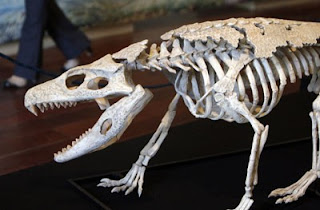
- Looking great overall, though the skull is probably too deep. As pointed out in this TetZoo [44], Kelenken has a very non-traditional, shallow skull, that may indicate most terror bird reconstructions with more massive skulls (including the skeletal you posted--sure that's of Kelenken? It lacks the humungo jugal bar, etc.) are in error. There's a pic of the skull on the site. As for the wings, I'd almost imagine the body plumage would hide them completely, like you see in most Moa recons. Do we know if they had quill knobs, etc. which would indicate large ostrich-like remiges for display? Dinoguy2 (talk) 05:26, 5 November 2009 (UTC)
- Hmm, the outline of that skeletal is used here at the bottom: http://scienceblogs.com/tetrapodzoology/2008/06/2007_year_of_terror_birds.php But yeah, looking at the skull, and this restoration[45], it probably shouldn't be so deep. How would that jugal bar affect the external appearance, and more specifically, this drawing? Is it wrong? FunkMonk (talk) 07:31, 5 November 2009 (UTC)
- Looking great overall, though the skull is probably too deep. As pointed out in this TetZoo [44], Kelenken has a very non-traditional, shallow skull, that may indicate most terror bird reconstructions with more massive skulls (including the skeletal you posted--sure that's of Kelenken? It lacks the humungo jugal bar, etc.) are in error. There's a pic of the skull on the site. As for the wings, I'd almost imagine the body plumage would hide them completely, like you see in most Moa recons. Do we know if they had quill knobs, etc. which would indicate large ostrich-like remiges for display? Dinoguy2 (talk) 05:26, 5 November 2009 (UTC)
- I'm not so sure how the wings should be depicted. It seems like the primary feathers would extend below the margin of the breast, which seems a little odd. But that's how it seems to be shown in John Conway's Andalgalornis, so I'd say that's your best bet. Your illustration looks great, by the way, especially the head! Smokeybjb (talk) 05:21, 5 November 2009 (UTC)
![[45]](https://home.earthlink.net/~rehrdc/producerwebsite/terrorbird_files/droppedImage.jpg)
How about this? [46] FunkMonk (talk) 15:49, 10 November 2009 (UTC)
![[46]](http://img697.imageshack.us/img697/9660/untitleaad1j.jpg)
- Much better, though the eye looks very large compared to the sclerotic ring in the skeletal and orbit in the skull photo. I'd also make the eye look as un-human like as possible--the Care Bear cheeks have the side effect of making any restoration come out a bit anthro, so restoring this guy in a way that looks like he's not intentionally smiling devilishly at you is tricky... A smaller, beadier eye will likely help. Dinoguy2 (talk) 16:56, 10 November 2009 (UTC)
- Yeah (only the dark part is the actual eyeball, the white around it is supposed to be skin), was going to make the eyes black, but then noted that seriemas (at least Cariama) have very different eyes.[47] Would it be within the possible to have that different eyes? Maybe some other, more related birds have even more different eyes? But I couldn't find a proper eye picture of that other Seriema genus, Chunga, so don't know how the eye looks, but it appears that it may well be black from the images I could find. By the way, from the Kelenken skull image, I was not really sure where the nostril and eye should be, does it look alright? The Knight/Burian head crest is quite unimaginative by the way, but had to make the skull longer, so let's say it's an homage... FunkMonk (talk) 20:11, 10 November 2009 (UTC)
- Ah, ok, so maybe it just did look like a smiling CareBare and there's nothing really to be done ;) No worries, if that's skin then it should look ok when colored. You're probably right, based on the skull it looks like the nostril should be farther forward, creating a 'kink' in the keratin beak near the dorsal side. Dinoguy2 (talk) 22:33, 10 November 2009 (UTC)
- Moved the nostril among other things, is it ok?: http://img694.imageshack.us/img694/5516/45007682.jpg FunkMonk (talk) 04:00, 11 November 2009 (UTC)
- Ah, ok, so maybe it just did look like a smiling CareBare and there's nothing really to be done ;) No worries, if that's skin then it should look ok when colored. You're probably right, based on the skull it looks like the nostril should be farther forward, creating a 'kink' in the keratin beak near the dorsal side. Dinoguy2 (talk) 22:33, 10 November 2009 (UTC)
- Yeah (only the dark part is the actual eyeball, the white around it is supposed to be skin), was going to make the eyes black, but then noted that seriemas (at least Cariama) have very different eyes.[47] Would it be within the possible to have that different eyes? Maybe some other, more related birds have even more different eyes? But I couldn't find a proper eye picture of that other Seriema genus, Chunga, so don't know how the eye looks, but it appears that it may well be black from the images I could find. By the way, from the Kelenken skull image, I was not really sure where the nostril and eye should be, does it look alright? The Knight/Burian head crest is quite unimaginative by the way, but had to make the skull longer, so let's say it's an homage... FunkMonk (talk) 20:11, 10 November 2009 (UTC)
![[47]](http://www.rosanevolpatto.trd.br/seriema2.jpg)

It's pretty much finished now, any thoughts? FunkMonk (talk) 16:47, 23 November 2009 (UTC)
Dalanistes edit
Someone else made and uploaded File:Dalanistes03.png, but it's clearly based on this Ambulocetus picture... Evercat (talk) 20:43, 9 November 2009 (UTC)
- I mentioned that to the uploader. He said he used that image as a model and used Bryce to alter it. I warned him that it was a derivative work of a copyrighted image that was unsuitable for upload to Commons. He uploaded a new version of Dalanistes that was somewhat different than the original (which has been deleted), but I still think it is similar enough to the Ambulocetus illustration to be considered a derivative work and thus should not be included in Wikipedia or Wikimedia Commons. Smokeybjb (talk) 21:06, 9 November 2009 (UTC)
- Yeah, that should obviously be deleted. If it was on Commons, I'd tag it as a copyvio, but I've never nominated anything for deletion on Wikipedia, it is a lot more complicated process. FunkMonk (talk) 21:29, 9 November 2009 (UTC)
- I'd say it qualifies for speedy deletion, specifically under F9: Unambiguous copyright infringement. That's how the last one was deleted. Smokeybjb (talk) 23:27, 9 November 2009 (UTC)
- Yeah, that should obviously be deleted. If it was on Commons, I'd tag it as a copyvio, but I've never nominated anything for deletion on Wikipedia, it is a lot more complicated process. FunkMonk (talk) 21:29, 9 November 2009 (UTC)
Odontogriphus edit
_drawing.png)
Here is an old image of mine depicting the pre-mollusk Odontogriphus, which got replaced with a diagrammatical illustration detailing this animals anatomy, probably due to some flaws. The one obvious flaw I detected was confusing the salivary glands for eyes. But should I make a new illustration, should the salivary glands be bulging out or completely internalized? Any other errors you see here? Giant Blue Anteater (talk) 22:44, 28 November 2009 (UTC)
- I barely know anything when it comes to invertebrates, especially Burgess fauna, but I'd say that the salivary glands would be internal. They're not visible in the restoration in Caron et al. (2006), which can be found here. Also, I'd suggest that you make the ends of the animal a bit rounder, based on the shape of the fossils. Smokeybjb (talk) 02:25, 29 November 2009 (UTC)
- Don't know much either, but I agree, it should definitely have less sharp edges. FunkMonk (talk) 21:07, 30 November 2009 (UTC)
- I wasn't even aware of this critter! If at all possible, I'd try to make your version less diagrammatic, with some more natural shading and maybe a more pinkish coloration. Honestly when I first saw your rendition I thought it was some kind of TV remote! I'd also be hard pressed to figure out that the image in the current article is an organic life form without the accompanying text. The article desperately needs a naturalistic reconstruction that looks more like a flatworm or slug than a garage door opener... Dinoguy2 (talk) 17:02, 1 December 2009 (UTC)
- What? I thought everyone who is interested in paleontology has at least read or heard the name Odontogriphus! Anyway, I am in the process of making a new image that dosen't look like a remote control device. Is there any evidence of segmentation or at least bands in this animal? Giant Blue Anteater (talk) 02:30, 3 December 2009 (UTC)
- Again, not that I'm the guy to ask, but a cursory read suggests it had 'wrinkles' near the anterior end. I wonder if these could be vestiges of segmentation in recent ancestors? Dinoguy2 (talk) 15:42, 3 December 2009 (UTC)
- Also, if you wish for a naturalistic illustration, the best I can do is one attached to a smooth rock, feeding on the algae that grew on it, with another individual in the background doing the undulating movement it is commonly portrayed doing, although I am beginning to think that such a feat is unlikely, considering that it had a semi-rigid 'shell' on its back. Would this 'shell' also account for a smooth body or would it remain "wrinkly"? Giant Blue Anteater (talk) 06:31, 4 December 2009 (UTC)
- Again, not that I'm the guy to ask, but a cursory read suggests it had 'wrinkles' near the anterior end. I wonder if these could be vestiges of segmentation in recent ancestors? Dinoguy2 (talk) 15:42, 3 December 2009 (UTC)
- What? I thought everyone who is interested in paleontology has at least read or heard the name Odontogriphus! Anyway, I am in the process of making a new image that dosen't look like a remote control device. Is there any evidence of segmentation or at least bands in this animal? Giant Blue Anteater (talk) 02:30, 3 December 2009 (UTC)
- I wasn't even aware of this critter! If at all possible, I'd try to make your version less diagrammatic, with some more natural shading and maybe a more pinkish coloration. Honestly when I first saw your rendition I thought it was some kind of TV remote! I'd also be hard pressed to figure out that the image in the current article is an organic life form without the accompanying text. The article desperately needs a naturalistic reconstruction that looks more like a flatworm or slug than a garage door opener... Dinoguy2 (talk) 17:02, 1 December 2009 (UTC)
- Don't know much either, but I agree, it should definitely have less sharp edges. FunkMonk (talk) 21:07, 30 November 2009 (UTC)
I've been wanting to do a ground sloth for a while, so here it is.[48] Based the proportions on the skeleton in the article, and external stuff on Choloepus. Most ground sloth restorations show visible ears for some reason, but I didn't add that, since both living sloth genera do not have such. Is there any evidense for visible pinnae? FunkMonk (talk) 21:12, 30 November 2009 (UTC)
![[48]](http://img263.imageshack.us/img263/4263/sloyhchhopy.jpg)
- According to User:Doc sloth, that skeleton was of Nothrotheriops. I'm not sure if the ears would have been visible, maybe he could help you out with that? Smokeybjb (talk) 22:00, 30 November 2009 (UTC)

- Oh yeah, I just forgot to rename the image of the skeleton, it was labeled as Nothrotherium where I found it... But yeah, I'll ask him! And here's a colour version. FunkMonk (talk) 22:15, 30 November 2009 (UTC)
- Looks great! The only thing I can think of (and I'm not sure that I'm correct) is that if the ears were not visible, there would be long, thick hair covering them. Perhaps the neck area of your sloth should be more thickly covered in long hair. You can see what I mean here[49]. However, I'm not sure how large ground sloth ears would have been relative to their skulls, and if their relative size is even comparable to that of tree sloths. Smokeybjb (talk) 21:11, 2 December 2009 (UTC)
- Oh yeah, I just forgot to rename the image of the skeleton, it was labeled as Nothrotherium where I found it... But yeah, I'll ask him! And here's a colour version. FunkMonk (talk) 22:15, 30 November 2009 (UTC)
![[49]](http://img23.imageshack.us/img23/4294/nothrotheriopssuggestioz.jpg)
Restoration[50] based on the skeletal in the new paper that came out a few days ago in the Zoological Journal of the Linnaean Society. See the Chinleana and Tetrapod Zoology posts for some of the images in the new paper. Unfortunately, I do not have access to the journal so I won't be able to read the full text article. If anyone has access to the journal, please speak up if you find that my reconstruction is inaccurate in any way! Smokeybjb (talk) 20:36, 2 December 2009 (UTC)
![[50]](http://img43.imageshack.us/img43/3539/vancleavea2.jpg)
- Looks cool, seems like the chin should be "weaker", and that the femur should be longer and the hip joint should be placed farther back, and the lower leg and arm should be thicker too, I've drawn it in here: [51] FunkMonk (talk) 23:09, 2 December 2009 (UTC)
![[51]](http://img694.imageshack.us/img694/3539/vancleavea2.jpg)
![[52]](http://img46.imageshack.us/img46/5972/vancleavea3.jpg)
![[53]](http://img80.imageshack.us/img80/5905/vancleavea3skeletalover.jpg)


I have made an image of the furcacaudiform thelodont, Sphenonectris. I used the following images as refferences: [54], [55], and [56]. What do you think? Giant Blue Anteater (talk) 02:32, 5 December 2009 (UTC)
![[54]](http://pwrmark.biology.ualberta.ca/ModelsandCasts/ModelsandCasts/images/sphenonectris_cast.GIF)
![[55]](http://fossils.valdosta.edu/fossil_pages/fossils_dev/images/f5.gif)
![[56]](http://www.palaeos.com/Vertebrates/Units/050Thelodonti/Images/Furcacaudiformes1.jpg)
- Looks good, but maybe the dorsal margin of the head should be somewhat concave rather than straight, like in the fossil and the restoration in Wilson & Caldwell (1998). Smokeybjb (talk) 15:37, 5 December 2009 (UTC)
- Yeah, and seems like the ventral side should be more bulgy, rather than straight too. By the way, you might want to create a Commons account, then you can upload your images there and all non-English Wikipedias will be able to use your images too. I'm hesitant to upload your images there myself, because then you won't be able to update them, unless you have an account there. FunkMonk (talk) 15:42, 5 December 2009 (UTC)
- I do have an account there, but I forgot my password. To make matters worse, I did not register my e-mail account there either. Giant Blue Anteater (talk) 16:54, 5 December 2009 (UTC)
- If you make a global account with a unified login, you won't need to make a new account for Commons, and your password should be the same there as for Wikipedia. Smokeybjb (talk) 17:04, 5 December 2009 (UTC)
- Yeah, that's what I did, changed the names of my other accounts to this one too. FunkMonk (talk) 17:18, 5 December 2009 (UTC)
- I will consider that in the future. Anyway, I have adjusted the shape of my reconstruction to fit your guys suggestions. Is it any better now? Giant Blue Anteater (talk) 17:28, 5 December 2009 (UTC)
- Yes, it's looking better. Based on the fossil, the caudal fin margin could have a serrated look, projecting out with each "ray". The restoration in Wilson & Caldwell (1998) has a smooth margin, so perhaps the serrated appearance in the fossil is due to partial decay before burial. Anyone know? Other than that, I don't have any other suggestions. Smokeybjb (talk) 22:05, 5 December 2009 (UTC)
- I'll say the serrated look of the caudal fin in the fossil was indeed due to decay, but perhaps... Giant Blue Anteater (talk) 23:14, 5 December 2009 (UTC)
- Yes, it's looking better. Based on the fossil, the caudal fin margin could have a serrated look, projecting out with each "ray". The restoration in Wilson & Caldwell (1998) has a smooth margin, so perhaps the serrated appearance in the fossil is due to partial decay before burial. Anyone know? Other than that, I don't have any other suggestions. Smokeybjb (talk) 22:05, 5 December 2009 (UTC)
- I will consider that in the future. Anyway, I have adjusted the shape of my reconstruction to fit your guys suggestions. Is it any better now? Giant Blue Anteater (talk) 17:28, 5 December 2009 (UTC)
- Yeah, that's what I did, changed the names of my other accounts to this one too. FunkMonk (talk) 17:18, 5 December 2009 (UTC)
- If you make a global account with a unified login, you won't need to make a new account for Commons, and your password should be the same there as for Wikipedia. Smokeybjb (talk) 17:04, 5 December 2009 (UTC)
- I do have an account there, but I forgot my password. To make matters worse, I did not register my e-mail account there either. Giant Blue Anteater (talk) 16:54, 5 December 2009 (UTC)
- Yeah, and seems like the ventral side should be more bulgy, rather than straight too. By the way, you might want to create a Commons account, then you can upload your images there and all non-English Wikipedias will be able to use your images too. I'm hesitant to upload your images there myself, because then you won't be able to update them, unless you have an account there. FunkMonk (talk) 15:42, 5 December 2009 (UTC)
Before I upload a new version to Wikipedia, what do you think of this ([57])? Adjustments include hightened body, fin rays shaped more like candy corn, and a concave mouth, as well as adding a margin at the base of the fin rays. Giant Blue Anteater (talk) 19:20, 8 January 2010 (UTC)
![[57]](http://img526.imageshack.us/img526/8448/sphenonectrisii.png)
- Excellent! The new version is definitely better than the previous one. Smokeybjb (talk) 22:08, 8 January 2010 (UTC)
- Great! Uploading away... Giant Blue Anteater (talk) 22:09, 8 January 2010 (UTC)
Very hard to find info on this one, so it might have been foolish to start it... But here is a sketch: [58] Used these images as reference: [59][60][61] Anyone have better images of the skeleton? FunkMonk (talk) 17:34, 5 December 2009 (UTC)
![[58]](http://img694.imageshack.us/img694/8062/alllrr2copy.jpg)
![[59]](https://en.wikipedia.org/wiki/File:Opetiosaurus.jpg)
![[60]](http://www.palaeos.com/Vertebrates/Units/Unit260/Images/Aigialosauridae1.jpg)
- Yep. There are some really great skeletals in "Aigialosaurs: Mid-Cretaceous varanoid lizards" (Journal of Vertebrate Paleontology 12(1):66-86). If you don't have access to the article, you can see the skeletals here. I sketched this creature a while back, and meant to put it up for review, but never got around to it. In aigialosaurs, the digits were not fused into paddles like they were in mosasaurs; in fact, the limbs look very similar to modern varanoids, and the unguals (yes, they were still there) have the same shape as those of terrestrial squamates.
- Keep in mind that although Opetiosaurus is a junior synonym of Aigialosaurus, its type species, O. bucchichi, is still its own species of Aigialosaurus. I don't know which species of Aigialosaurus the Palaeos skeleton belongs to, it may be A. dalmaticus. If this is the case, your restoration would be sort of like a chimaera. Looking at the skeletals, it seems as though A. dalmaticus has longer hind limbs, while in A. bucchichi the fore and hind limbs are roughly equal in length. Therefore, although the head of your restoration is based on the skull of A. bucchichi, the body looks more like A. dalmaticus as it has longer hind limbs. Smokeybjb (talk) 21:48, 5 December 2009 (UTC)
- Thanks for that! Is your own version ready for review? What species did you draw? Then we could maybe have an image of each species. FunkMonk (talk) 22:01, 5 December 2009 (UTC)
- Good idea. Mine's of A. dalmaticus. I haven't quite finished the sketch, but I'll put it up for review as soon as it's ready. Also, it seems like the tail should be a bit less fusiform in your restoration. Usually, the tails of mosasauroids are the same depth throughout their length, except in some genera such as Clidastes and Plotosaurus, where the neural spines are more elongate in some sections. Smokeybjb (talk) 02:22, 6 December 2009 (UTC)
- Perfect then, we made a differnet species each anyway (or well, the head of mine at least)! Yeah, I'll fix the tail too, was mislead by the mount where most of the tail is out of frame. FunkMonk (talk) 02:36, 6 December 2009 (UTC)
- Here it is, A. dalmaticus[62]. The pose is somewhat boring compared to yours :). Smokeybjb (talk) 20:52, 6 December 2009 (UTC)
- I like your pose! And damn, seems like I'll have to remake everything but the head... Will make the bodyline more curved to the side.FunkMonk (talk) 21:42, 6 December 2009 (UTC)
- I'd say you'd just have to make the flippers into arms and legs and readjust the size of the hindlimbs, but hey, do whatever you want! Smokeybjb (talk) 22:34, 6 December 2009 (UTC)
- Changed some stuff now, how about it? http://img156.imageshack.us/img156/7013/ufgy.jpg FunkMonk (talk) 04:53, 5 January 2010 (UTC)
- It's looking great so far, but, looking at the skeletal, the hands and feet could be just a bit smaller relative to the length of the limbs. Also, the chest should probably be less deep, as it appears that the coracoid is closer to the vertebrae in A. buccichi than in A. dalmaticus. Do you see any innacuracies in my restoration, by the way? Smokeybjb (talk) 04:21, 9 January 2010 (UTC)
- Changed some stuff now, how about it? http://img156.imageshack.us/img156/7013/ufgy.jpg FunkMonk (talk) 04:53, 5 January 2010 (UTC)
- I'd say you'd just have to make the flippers into arms and legs and readjust the size of the hindlimbs, but hey, do whatever you want! Smokeybjb (talk) 22:34, 6 December 2009 (UTC)
- I like your pose! And damn, seems like I'll have to remake everything but the head... Will make the bodyline more curved to the side.FunkMonk (talk) 21:42, 6 December 2009 (UTC)
- Here it is, A. dalmaticus[62]. The pose is somewhat boring compared to yours :). Smokeybjb (talk) 20:52, 6 December 2009 (UTC)
- Perfect then, we made a differnet species each anyway (or well, the head of mine at least)! Yeah, I'll fix the tail too, was mislead by the mount where most of the tail is out of frame. FunkMonk (talk) 02:36, 6 December 2009 (UTC)
- Good idea. Mine's of A. dalmaticus. I haven't quite finished the sketch, but I'll put it up for review as soon as it's ready. Also, it seems like the tail should be a bit less fusiform in your restoration. Usually, the tails of mosasauroids are the same depth throughout their length, except in some genera such as Clidastes and Plotosaurus, where the neural spines are more elongate in some sections. Smokeybjb (talk) 02:22, 6 December 2009 (UTC)
- Thanks for that! Is your own version ready for review? What species did you draw? Then we could maybe have an image of each species. FunkMonk (talk) 22:01, 5 December 2009 (UTC)
![[62]](http://img687.imageshack.us/img687/63/adalmaticus.jpg)
![[63]](http://img246.imageshack.us/img246/7013/ufgy.jpg)
![[64]](http://img41.imageshack.us/img41/63/adalmaticus.jpg)


Rather than immediately uploading my illustrations here, I'm just going to upload them to Imageshack until I unify my accounts. Here is my image of yet another thelodont, Furcacauda: [65], using the following two references: [66] and [67]. There is already an image of Furcacauda on the article though, by Stanton Fink. But is this image still worthy of inclusion? Giant Blue Anteater (talk) 00:36, 6 December 2009 (UTC)
![[65]](http://img69.imageshack.us/img69/5111/furcacauda.png)
![[66]](http://pwrmark.biology.ualberta.ca/ModelsandCasts/ModelsandCasts/images/Cast.Fork-tailed.thelodont.GIF)
![[67]](http://users.atw.hu/fishindex/images/vertebrata/thelodus.gif)
- Even if there is already an image in an article, any image is worthy of inclusion so long as it is accurate! The only thing I can think of is that the fin tips should project farther from the animal, like in the restoration you used as reference. By the way, the url for that reference says Thelodus. The webpage it came from[68] doesn't label the illustration. It looks like Furcacauda, but are you sure that it is the genus? Smokeybjb (talk) 02:54, 6 December 2009 (UTC)
- The fossil dosen't seem to bear evidence for long caudal fin lobes, so the illustration I took inspiration from probably had them for artistic license. Also, there are smaller versions of this picture in various websites (Palaeos.com included). As for the filename, the fish portrayed is indeed a Furcacauda. I do not know the original source of that illustration, but I can tell you that is not a Thelodus, as it looked a lot different. Giant Blue Anteater (talk) 05:37, 6 December 2009 (UTC)
- I don't know much about thelodonts, so I didn't really know what Thelodus looked like. All the fossil photographs of Furcacauda that I can find on the web are pretty low-resolution, so it's hard to tell how far the caudal lobes extend. I'll have to take your word for it. By the way, what species have you restored? I assume that it's F. fredholmae due to the shark-like dorsal fin. In F. heintzae, there is a supplementary ventral lobe under the main ventral lobe of the caudal fin, in case you are restoring that species. It seems that the existig illustration in the article doesn't depict that, though. Also, Wilson & Caldwell (1998) say that F. heintzae bears a pair of "fin-flaps" on its trunk, which the existing restoration doesn't show either. Smokeybjb (talk) 15:33, 6 December 2009 (UTC)
- I did not have any particular species in mind while drawing this, just the genus. Is that a problem? By "fin-flaps", you mean lateral fins, right? Giant Blue Anteater (talk) 21:41, 6 December 2009 (UTC)
- Yeah, I guess you could say that. Wilson & Caldwell (1998) seemed to suggest that they weren't really fins per se but rather fin-like appendages. These fins didn't extend from the midline like in Shielia, but rather from the bottom of the trunk near the ventral margin, sort of like this. The reason why I asked you what species it is is because the two species appear quite different, and you shouldn't combine diferent features of the two into one animal. Smokeybjb (talk) 22:12, 6 December 2009 (UTC)
- I'm making a new illustration based on the one I made to be an F. heintzae. From my understanding of your description of the supplementary ventral lobe, you mean that a pelvic fin-like structure should be placed where the ventral skid is? And F. heintzae lacked the dorsal fin? But if the illustration I made would be any species, I guess it would be an F. fredholmae, but any more changes I should make? Giant Blue Anteater (talk) 02:44, 12 December 2009 (UTC)
- Yes, the lobes should be placed at the ventral skid. There is a pair of them, but I guess in lateral view the right lobe would be hidden behind the left one. In the restoration in Wilson & Caldwell (1998), the lobes project ventrally rather than laterally, like in the example I showed you. And one more thing, the lobes shouldn't be as thin as the dorsal fin, but more like fleshy flaps. Therefore, I suggest you extend the light gray areas of the restoration rather than making the lobes bluish like the other fins. F. heintzae did not lack a dorsal fin, it was just more swept back and less "shark-like" than that of F. fredholmae. I don't see any changes you should make for F. fredholmae, so you might as well label your original illustration as that species and put it in the article. And if you are restoring F. heintzae in your new illustration, that would allow us to have illustrations of each species in the article. Smokeybjb (talk) 17:02, 12 December 2009 (UTC)
- I have now uploaded a modified version of my illustration for F. fredholmae. As for F. heintzae, do you consider this of improvement? [69] Giant Blue Anteater (talk) 07:53, 13 December 2009 (UTC)
- Yes, it looks good! Nice color choice with F. fredholmae, too. But one more thing before you upload F. heintzae: I mistakenly was talking about the fin flaps after you asked about the ventral lobe. The lobe would be part of the caudal fin projecting from the ventral skid, like you thought. It would project from below the posterior crescent margin of the fin, like this.[70] If the main caudal lobes were longer like in the restoration used as reference, then the supplementary lobe would end before the tip of the lower lobe, but since you do not think they were that long, the supplementary lobe would probably end with the lower lobe.
- Alright, I have added that little lobe (here: [71]). Should I keep the membrane I added between the main lobe and the peculiar "pinky" lobe or remove it? Giant Blue Anteater (talk) 19:35, 13 December 2009 (UTC)
- No, there doesn't seem to be any evidence of a membrane between the two lobes.Smokeybjb (talk) 00:37, 14 December 2009 (UTC)
- I have now uploaded the finished project without the membrane between the little lobe and the big lobe. Any further amendments to be made? By the way, do you know anything about Cometicercus? I cannot find much discriptive information about it on the Internet, but the only restoration I could find is Stanton Fink's, which can be seen here: [72]. Giant Blue Anteater (talk) 02:39, 14 December 2009 (UTC)
- Awesome! They look great now, I don't see any other inaccuracies. Cometicercus is known from only one specimen, and the front half of the body is not preserved, so restorations would be a bit speculative. Here's a photo from Wilson & Caldwell (1998).[73] Unfortunately, I don't know of any other good life restorations that you could use for reference. There's one in Wilson & Caldwell, but it's not shown in lateral view. The lower lobe of the caudal fin seems much longer than the upper lobe, though. Smokeybjb (talk) 02:37, 17 December 2009 (UTC)
- I have now uploaded the finished project without the membrane between the little lobe and the big lobe. Any further amendments to be made? By the way, do you know anything about Cometicercus? I cannot find much discriptive information about it on the Internet, but the only restoration I could find is Stanton Fink's, which can be seen here: [72]. Giant Blue Anteater (talk) 02:39, 14 December 2009 (UTC)
- No, there doesn't seem to be any evidence of a membrane between the two lobes.Smokeybjb (talk) 00:37, 14 December 2009 (UTC)
- Alright, I have added that little lobe (here: [71]). Should I keep the membrane I added between the main lobe and the peculiar "pinky" lobe or remove it? Giant Blue Anteater (talk) 19:35, 13 December 2009 (UTC)
- Yes, it looks good! Nice color choice with F. fredholmae, too. But one more thing before you upload F. heintzae: I mistakenly was talking about the fin flaps after you asked about the ventral lobe. The lobe would be part of the caudal fin projecting from the ventral skid, like you thought. It would project from below the posterior crescent margin of the fin, like this.[70] If the main caudal lobes were longer like in the restoration used as reference, then the supplementary lobe would end before the tip of the lower lobe, but since you do not think they were that long, the supplementary lobe would probably end with the lower lobe.
- I have now uploaded a modified version of my illustration for F. fredholmae. As for F. heintzae, do you consider this of improvement? [69] Giant Blue Anteater (talk) 07:53, 13 December 2009 (UTC)
- Yes, the lobes should be placed at the ventral skid. There is a pair of them, but I guess in lateral view the right lobe would be hidden behind the left one. In the restoration in Wilson & Caldwell (1998), the lobes project ventrally rather than laterally, like in the example I showed you. And one more thing, the lobes shouldn't be as thin as the dorsal fin, but more like fleshy flaps. Therefore, I suggest you extend the light gray areas of the restoration rather than making the lobes bluish like the other fins. F. heintzae did not lack a dorsal fin, it was just more swept back and less "shark-like" than that of F. fredholmae. I don't see any changes you should make for F. fredholmae, so you might as well label your original illustration as that species and put it in the article. And if you are restoring F. heintzae in your new illustration, that would allow us to have illustrations of each species in the article. Smokeybjb (talk) 17:02, 12 December 2009 (UTC)
- I'm making a new illustration based on the one I made to be an F. heintzae. From my understanding of your description of the supplementary ventral lobe, you mean that a pelvic fin-like structure should be placed where the ventral skid is? And F. heintzae lacked the dorsal fin? But if the illustration I made would be any species, I guess it would be an F. fredholmae, but any more changes I should make? Giant Blue Anteater (talk) 02:44, 12 December 2009 (UTC)
- Yeah, I guess you could say that. Wilson & Caldwell (1998) seemed to suggest that they weren't really fins per se but rather fin-like appendages. These fins didn't extend from the midline like in Shielia, but rather from the bottom of the trunk near the ventral margin, sort of like this. The reason why I asked you what species it is is because the two species appear quite different, and you shouldn't combine diferent features of the two into one animal. Smokeybjb (talk) 22:12, 6 December 2009 (UTC)
- I did not have any particular species in mind while drawing this, just the genus. Is that a problem? By "fin-flaps", you mean lateral fins, right? Giant Blue Anteater (talk) 21:41, 6 December 2009 (UTC)
- I don't know much about thelodonts, so I didn't really know what Thelodus looked like. All the fossil photographs of Furcacauda that I can find on the web are pretty low-resolution, so it's hard to tell how far the caudal lobes extend. I'll have to take your word for it. By the way, what species have you restored? I assume that it's F. fredholmae due to the shark-like dorsal fin. In F. heintzae, there is a supplementary ventral lobe under the main ventral lobe of the caudal fin, in case you are restoring that species. It seems that the existig illustration in the article doesn't depict that, though. Also, Wilson & Caldwell (1998) say that F. heintzae bears a pair of "fin-flaps" on its trunk, which the existing restoration doesn't show either. Smokeybjb (talk) 15:33, 6 December 2009 (UTC)
- The fossil dosen't seem to bear evidence for long caudal fin lobes, so the illustration I took inspiration from probably had them for artistic license. Also, there are smaller versions of this picture in various websites (Palaeos.com included). As for the filename, the fish portrayed is indeed a Furcacauda. I do not know the original source of that illustration, but I can tell you that is not a Thelodus, as it looked a lot different. Giant Blue Anteater (talk) 05:37, 6 December 2009 (UTC)
![[69]](http://img138.imageshack.us/img138/3360/furcacaudaheintzae.png)
![[70]](http://img31.imageshack.us/img31/7932/fheintzae.png)
![[71]](http://img199.imageshack.us/img199/3360/furcacaudaheintzae.png)
![[73]](http://img194.imageshack.us/img194/7264/cometicercustalimaaae.jpg)
Here's a change from life restorations. I've made a diagram of aetosaur osteoderms.[74] It was made as an SVG file using Inkscape, so it will be easy to fix any problems. It's based on a skeletal of Neoaetosauroides from an article in the journal Ameghiniana.[75] An introduction to aetosaur osteoderms can be found in this Chinleana blog post.[76] Have I labeled anything wrong, or are there any anatomical inaccuracies or aesthetic problems? I don't know how to upload SVGs using other sites, so you can scrutinize the diagram in greater detail once I upload it to Commons. I haven't labeled the osteoderms on the throat and pelvic regions yet because I do not know what the technical terms are for them. Anyone know? I may eventually make another diagram labeling the morphology of individual aetosaur osteoderms, too. Smokeybjb (talk) 19:38, 12 December 2009 (UTC)
![[74]](http://img193.imageshack.us/img193/2985/aetosaur.png)

- Alright, here's the uploaded image. I guess I can just upload a new version if I find out the names for the throat and pelvic osteoderms. Smokeybjb (talk) 03:51, 9 January 2010 (UTC)
- Yup. It's nice! FunkMonk (talk) 05:05, 9 January 2010 (UTC)
- You migh want to take a look at this new Typothorax paper, it has a similar diagram: http://www.informaworld.com/smpp/content~db=all~content=a922419027~frm=titlelink FunkMonk (talk) 14:28, 28 May 2010 (UTC)
- Yup. It's nice! FunkMonk (talk) 05:05, 9 January 2010 (UTC)
- Alright, here's the uploaded image. I guess I can just upload a new version if I find out the names for the throat and pelvic osteoderms. Smokeybjb (talk) 03:51, 9 January 2010 (UTC)

Not a lot is known about this guy, but thanks to Smokeybjb, he has fished an photograph of the fossil, which is rather largely incomplete. So incomplete that it can only be speculated what the rest of the body looked like. Which shape is more likely for Cometicercus? [77] Also, even though Wilson and Caldwell's restoration is not a lateral view, may I see it anyway? Giant Blue Anteater (talk) 02:08, 18 December 2009 (UTC)
![[77]](http://img684.imageshack.us/img684/9611/cometocercus.png)
- Of course! Here it is (Cometicercus is "a" in the illustration).[78] Although the entire body is not shown in lateral view, the anterior half sort of is, so it could be a good reference for your restoration. (Ignore what I previously said; I misread the caption and thought Cometicercus was something else!) I would say the upper outline in your line drawing is more likely, as the ventral margin in most furcacaudiforms was either relatively straight or slightly concave. Also, judging by the fossil, the trunk region seems to extend downwards in a sort of "keel" as it does in Furcacauda with a pronounced anal notch. There is definitely a dorsal fin too, even though Stanton Fink's illustration lacks it. Smokeybjb (talk) 03:53, 20 December 2009 (UTC)
- Oh crappie. I already was drawing the bottom one and in a shape similar to Stanton's. Well, good thing I saved the line drawing for the one with the hump-backed bottom-feeder pose. And, if you know what, I actually kind of like the Furcacauda-like shape of Caldwell's illustration better. Also... I think there are problems to be corrected in all of my furcacaudiform illustrations. I am now actually displeased at how the pharyngeal slits are sloping downward rather than being organized straightly. Also, should I make the mouths concave or are they fine the way they are? Also, I think I added two few fin rays (6), when according to W. & C., there were 8-14 of them. By the way, what is "b"? Is it Drepanolepis or Pezopallichthys? Giant Blue Anteater (talk) 07:21, 20 December 2009 (UTC)
- And here is my work so far: [79] Giant Blue Anteater (talk) 23:22, 21 December 2009 (UTC)
- "b" is Drepanolepis. According to Wilson & Caldwell, Furcacauda heintzae had eight intermediate lobes (fin rays) on the caudal fin and F. fredholmae had at least ten. They also said that Cometicercus probably had ten intermediate lobes. There are four intermediate lobes seen on the dorsal primary lobe, but it is likely that there was five because other furcacaudids have an equal number of intermediate lobes on each primary lobe, and five intermediate lobes are seen on the ventral primary lobe of Cometicercus. Most restorations of furcacaudids have the mouth slightly concave, but I do not know for sure if this is always the case. In the holotype of Sphenonectris where the oral region is best preserved, the mouth seems to be very concave. You should probably add this concavity to your Sphenonectris restoration. Also, the intermediate lobes of the caudal fin look a bit too thin and ray-like in your restorations, especially for Sphenonectris. Perhaps if you widened the base of each lobe they would appear less ray-like. Smokeybjb (talk) 21:27, 23 December 2009 (UTC)
- I'll get to work on my other furcacadiformes. But what do you think of the final version of Cometicercus I just uploaded to Wikipedia? Also, could you get me images (fossils or otherwise) of the following: Drepanolepis, Pezopallichthys? Sorry, I do not have access to Wilson & Caldwell's paper, so I am requesting them from you. Giant Blue Anteater (talk) 22:24, 7 January 2010 (UTC)
- Anyone up for creating a furcacadiformes article now we're at it? I don't know anything about them, so I can't really do it. FunkMonk (talk) 16:14, 9 January 2010 (UTC)
- It looks good, but maybe the dorsal fin could be wider, occupying more of the dorsal margin. That's what it's like in the fossil and the restoration. Here's Drepanolepis[80] and Pezopallichthys[81] from Wilson & Caldwell (I don't know of any other images). In the Pezopallichthys image, "a" shows left lateral view, "b" right lateral view (anterior half), "c" right lateral view of branchial row at lower right and caudal peduncle at left, "d" right lateral view of head and branchial region, "e" left lateral view of caudal peduncle and caudal lobes, "f" left lateral view of branchial region with stomach endocast at right, "g" left lateral view with branchial region below and mid-dorsal body margin at top, and "h" left lateral view showing details of branchial bars. For a Furcacadiformes article, we could use the new furcacaudiform restorations, and I could use Wilson & Caldwell as a reference. Does anyone know of any other good papers on furcacaudiforms? Smokeybjb (talk) 18:32, 9 January 2010 (UTC)
- Now those two are gonna be fun! Especially Pezopallichthys! As for the dorsal fin of Cometicercus, I will adjust it to your suggestion soon after I post the outline of my Drepanolepis illustration. Also, for the Furcacaudiformes article, maybe we could transfer some of the content from the Furcacauda article? Giant Blue Anteater (talk) 21:53, 9 January 2010 (UTC)
- And I have uploaded a new version with a widened dorsal fin. Giant Blue Anteater (talk) 04:42, 10 January 2010 (UTC)
- It looks good, but maybe the dorsal fin could be wider, occupying more of the dorsal margin. That's what it's like in the fossil and the restoration. Here's Drepanolepis[80] and Pezopallichthys[81] from Wilson & Caldwell (I don't know of any other images). In the Pezopallichthys image, "a" shows left lateral view, "b" right lateral view (anterior half), "c" right lateral view of branchial row at lower right and caudal peduncle at left, "d" right lateral view of head and branchial region, "e" left lateral view of caudal peduncle and caudal lobes, "f" left lateral view of branchial region with stomach endocast at right, "g" left lateral view with branchial region below and mid-dorsal body margin at top, and "h" left lateral view showing details of branchial bars. For a Furcacadiformes article, we could use the new furcacaudiform restorations, and I could use Wilson & Caldwell as a reference. Does anyone know of any other good papers on furcacaudiforms? Smokeybjb (talk) 18:32, 9 January 2010 (UTC)
- Anyone up for creating a furcacadiformes article now we're at it? I don't know anything about them, so I can't really do it. FunkMonk (talk) 16:14, 9 January 2010 (UTC)
- I'll get to work on my other furcacadiformes. But what do you think of the final version of Cometicercus I just uploaded to Wikipedia? Also, could you get me images (fossils or otherwise) of the following: Drepanolepis, Pezopallichthys? Sorry, I do not have access to Wilson & Caldwell's paper, so I am requesting them from you. Giant Blue Anteater (talk) 22:24, 7 January 2010 (UTC)
- "b" is Drepanolepis. According to Wilson & Caldwell, Furcacauda heintzae had eight intermediate lobes (fin rays) on the caudal fin and F. fredholmae had at least ten. They also said that Cometicercus probably had ten intermediate lobes. There are four intermediate lobes seen on the dorsal primary lobe, but it is likely that there was five because other furcacaudids have an equal number of intermediate lobes on each primary lobe, and five intermediate lobes are seen on the ventral primary lobe of Cometicercus. Most restorations of furcacaudids have the mouth slightly concave, but I do not know for sure if this is always the case. In the holotype of Sphenonectris where the oral region is best preserved, the mouth seems to be very concave. You should probably add this concavity to your Sphenonectris restoration. Also, the intermediate lobes of the caudal fin look a bit too thin and ray-like in your restorations, especially for Sphenonectris. Perhaps if you widened the base of each lobe they would appear less ray-like. Smokeybjb (talk) 21:27, 23 December 2009 (UTC)
![[78]](http://img189.imageshack.us/img189/733/fig10.jpg)
![[79]](http://img704.imageshack.us/img704/9611/cometocercus.png)
![[80]](http://img682.imageshack.us/img682/687/wc1.png)
![[81]](http://img294.imageshack.us/img294/4953/wc2.png)
Here's a sketch of the newly described notosuchian Yacarerani [82] based on this figure[83] and the first photograph in this news article[84]. Any problems? I don't have access to the Journal of Vertebrate Paleontology article that it was described in, so the postcranial portion of the restoration is just a generalized notosuchian body. Can someone who has access to the article please provide me with an image of the postcranial remains (if there is one) to use as reference, or tell how I can improve my restoration to be more accurate? Smokeybjb (talk) 19:59, 26 December 2009 (UTC)
![[82]](http://img692.imageshack.us/img692/6476/yacarerani.jpg)
![[83]](http://i49.tinypic.com/kdsha0.jpg)
- There were some diagrams posted on the Dinoforum, but they've since moved, but I'll see if I can find them when/if the thread is moved... But looks good so far! The eye seems to fill out the entire socket, but since crocodiles don't have sclerotic rings, I'm not sure how large their eyes would be. Maybe it's something to look into, if you don't know already. FunkMonk (talk) 17:10, 27 December 2009 (UTC)
- Some small crocodylians, especially juveniles, have large eyes that occupy much of the socket. Given that Yacarerani was very small, I suspect that its eyes filled most of the sockets and were quite large. I've made the eye in my restoration a bit smaller now[85], because it did seem a little too large in the socket to begin with... perhaps it should have had more fleshy lids around it to fill up some space. Smokeybjb (talk) 03:36, 31 December 2009 (UTC)
- Speaking of notosuchians, I've made an attempt at correcting ArthurWeasley's Anatosuchus[86], since he's not as active as he used to be and may not upload a new version. How does it look? Smokeybjb (talk) 18:52, 2 January 2010 (UTC)
- Both look nice! I removed that Anatosuchus image exactly because of the former legs, seems like it could be put back in now. FunkMonk (talk) 05:04, 9 January 2010 (UTC)
- Any luck with the diagrams? I'm not too keen on making an image that is based primarily on speculation when much of the material is already known. Smokeybjb (talk) 20:06, 12 January 2010 (UTC)
- I aksed, but seems like the old forum will stay closed... FunkMonk (talk) 20:47, 12 January 2010 (UTC)
- Oh well, I'm thinking of uploading a finished version of just the head, at least until I manage to get access to the JVP article or any of its figures. Smokeybjb (talk) 02:39, 19 January 2010 (UTC)
- I aksed, but seems like the old forum will stay closed... FunkMonk (talk) 20:47, 12 January 2010 (UTC)
- Any luck with the diagrams? I'm not too keen on making an image that is based primarily on speculation when much of the material is already known. Smokeybjb (talk) 20:06, 12 January 2010 (UTC)
- Both look nice! I removed that Anatosuchus image exactly because of the former legs, seems like it could be put back in now. FunkMonk (talk) 05:04, 9 January 2010 (UTC)
- Speaking of notosuchians, I've made an attempt at correcting ArthurWeasley's Anatosuchus[86], since he's not as active as he used to be and may not upload a new version. How does it look? Smokeybjb (talk) 18:52, 2 January 2010 (UTC)
- Some small crocodylians, especially juveniles, have large eyes that occupy much of the socket. Given that Yacarerani was very small, I suspect that its eyes filled most of the sockets and were quite large. I've made the eye in my restoration a bit smaller now[85], because it did seem a little too large in the socket to begin with... perhaps it should have had more fleshy lids around it to fill up some space. Smokeybjb (talk) 03:36, 31 December 2009 (UTC)
![[85]](http://img705.imageshack.us/img705/6476/yacarerani.jpg)
![[86]](http://img686.imageshack.us/img686/6416/anatosuchusbw2.jpg)
