| Image |
Article |
Description
|
 |
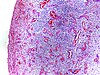
Neutrophils |
Micrograph showing neutrophils in acute inflammation.
|
 |
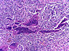
Neutrophils |
Micrograph showing neutrophil margination and extravasation in acute inflammation.
|
 |
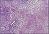
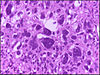
Plasma cells |
Plasma cell showing eccentrically placed round nucleus with cart wheel like chromatin.
|
 |
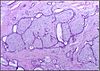
Eosinophils |
Micrograph showing low power view of Eosinophils.
|
 |
Eosinophils |
Micrograph showing high power view of Eosinophils. H&E stain.
|
 |
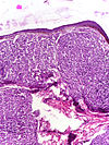
Lymphocytes |
Micrograph showing lymphocytes in caseating granuloma.
|
 |
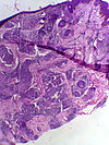
Fibroblasts |
|
|
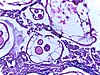
Foam cells |
|
 |
Hemosiderin laden macrophages |
Lung showing congestion with plenty of alveolar macrophages containing phagocytosed brownish granular hemosiderin pigment.
|
 |
Hemosiderin laden macrophages |
Lung showing congestion with plenty of alveolar macrophages containing phagocytosed brownish granular hemosiderin pigment.
|
|
Langhan cells |
|
 |
Foreign body giant cells |
Micrograph showing a foreign body engulfed by a giant cell.
|
 |
Granulation tissue |
Micrograph showing proliferating capillaries, fibroblasts and acute inflammatory cells.
|
 |
Granulation tissue |
Micrograph showing proliferating capillaries, fibroblasts and acute inflammatory cells.
|
 |
Foreign body granuloma |
Granulomatous reaction to keratin characterized by foreign body giant cells and chronic inflammatory cells.
|
 |
Foreign body granuloma |
Granulomatous reaction to keratin characterized by foreign body giant cells and chronic inflammatory cells.
|
|
Tuberculous granuloma |
Caseating granulomatous lesion with areas of amorphous granular eosinophilic necrotic debris known as caseation (on the right half) bordered by collections of epitheloid cells, Langhan giant cells and lymphocytes.
|
 |
Tuberculous granuloma |
Caseating granulomatous lesion with areas of amorphous granular eosinophilic necrotic debris known as caseation (on the right half) bordered by collections of epitheloid cells, Langhan giant cells and lymphocytes.
|
 |
Fat necrosis |
Breast lump showing an area of fat necrosis showing shadowy outlines of necrotic adipocytes surrounded by an inflammatory reaction with cholesterol clefts.
|
 |
Fat necrosis |
Breast lump showing an area of fat necrosis showing shadowy outlines of necrotic adipocytes surrounded by an inflammatory reaction with cholesterol clefts.
|
 |
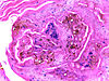
Calcinosis cutis |
Multiple amorphous basophilic calcium deposits in the dermis.
|
 |
Hemochromatosis liver |
Micrograph of hemochromatosis liver showing hepatocytes with coarse golden yellow granules of hemosiderin within the cytoplasm. These granules stain with Prussian blue stain.
|
|
Caseous necrosis |
Caseating granulomatous lesion with areas of amorphous granular eosinophilic necrotic debris known as caseation (on the right half) bordered by collections of epitheloid cells, Langhan giant cells and lymphocytes.
|
|
Caseous necrosis |
Caseating granulomatous lesion with areas of amorphous granular eosinophilic necrotic debris known as caseation (on the right half) bordered by collections of epitheloid cells, Langhan giant cells and lymphocytes.
|
 |
Fatty change in liver |
Hepatic parenchymal cell cytoplasm containing clear vacuoles containing fat of varying sizes, displacing the nucleus towards the periphery.
|
 |
Fatty change in liver |
Hepatic parenchymal cell cytoplasm containing clear vacuoles containing fat of varying sizes, displacing the nucleus towards the periphery.
|
 |
Carcinoma in situ |
CIN-III (HSIL) showing diffuse severe atypia with loss of maturation involving full thickness of epithelium with intact basement membrane.
|
 |
Carcinoma in situ |
CIN-I (LSIL) showing koilocytic atypia and intact basement membrane.
|
 |
Carcinoma in situ |
CIN-I (Low Grade Squamous Intraepithelial Lesion-LSIL) showing koilocytic atypia and intact basement membrane.
|
 |
Fluid cytology smear |
High power view showing intracytoplasmic mucin within malignant cells(Papanicolaou, 400X)
|
 |
Fluid cytology smear |
Low power view showing malignant cells arranged in glandular pattern (Papanicolaou, 100X)
|
|
Lymph node metastasis |
|
 |
Fibroadenoma breast |
A well circumscribed lesion showing proliferation of intralobular stroma compressing and distorting the epithelium.
|
 |
Fibroadenoma breast |
A well circumscribed lesion showing proliferation of intralobular stroma compressing and distorting the epithelium.
|
 |
Tumor giant cell |
Malignant neoplasm showing marked anaplasia. Note the marked nuclear pleomorphism, bizarre cells and tumor giant cells.
|
|
Capillary hemangioma |
|
 |
Lepromatous leprosy |
Skin biopsy showing epidermal atrophy and multiple dermal infiltrates.
|
 |
Lepromatous leprosy |
Infiltrate of foamy histiocytes with scarcity of lymphocytes in lepromatous leprosy.
|
 |
Lepromatous leprosy |
Skin biopsy showing epidermal atrophy and multiple dermal infiltrates.
|
 |
Lepromatous leprosy |
Skin biopsy showing epidermal atrophy and multiple inflammatory dermal infiltrates.
|
 |
Lepromatous leprosy |
Modified AFB staining in a case of lepromatous leprosy showing numerous rod shaped acid fast bacilli.
|
 |
Lepromatous leprosy |
Infiltrate of foamy histiocytes with scarcity of lymphocytes in lepromatous leprosy.
|
 |
Lepromatous leprosy |
Skin biopsy showing epidermal atrophy and multiple dermal infiltrates composed of mainly foamy macrophages.
|
 |
Lepromatous leprosy |
Infiltrate of foamy histiocytes with scarcity of lymphocytes in lepromatous leprosy.
|
 |
Lepromatous leprosy |
Infiltrate of foamy histiocytes with scarcity of lymphocytes in lepromatous leprosy.
|
 |
Lepromatous leprosy |
Skin biopsy showing subepidermal Grenz zone.
|
 |
Tuberculoid leprosy |
Skin biopsy showing multiple peri-appendageal granulomas.
|
 |
Tuberculoid leprosy |
Granulomatous lesion with epithelioid cells, Langhans giant cells and prominent lymphocyte infiltrate.
|
 |
Tuberculoid leprosy |
Granulomatous lesion with epithelioid cells, Langhans giant cells and prominent lymphocyte infiltrate.
|
 |
Tuberculoid leprosy |
Skin biopsy specimen showing multiple granulomas.
|
|
Tuberculous lymph node |
Caseating granulomatous lesion with areas of amorphous granular eosinophilic necrotic debris known as caseation (on the right half) bordered by collections of epitheloid cells, Langhan giant cells and lymphocytes.
|
|
Tuberculous lymph node |
Caseating granulomatous lesion with areas of amorphous granular eosinophilic necrotic debris known as caseation (on the right half) bordered by collections of epitheloid cells, Langhan giant cells and lymphocytes.
|
|
Tuberculous intestine |
|
 |
Helminth in appendix |
Micrograph showing lumen of appendix and cut section of pin worm.
|
 |
Chromoblastomycosis |
Micrograph of chromoblastomycosis showing sclerotic bodies in dermis.
|
 |
Amoebiasis in intestine |
Colonic biopsy showing trophozoite of Entamoeba Histolytica with ingested red blood cells.
|
 |
Amoebiasis in intestine |
Colonic biopsy showing trophozoite of Entamoeba Histolytica with ingested red blood cells.
|
|
Rhinosporidiosis |
|
 |
Filarial lymphadenitis |
Micrograph showing section of an adult filarial worm. Surrounding tissue shows dense infiltration of eosinophils.
|
|
Reactive hyperplasia of lymph node |
|
|
Diffuse B-cell lymphoma |
Caseating granulomatous lesion with areas of amorphous granular eosinophilic necrotic debris known as caseation (on the right half) bordered by collections of epitheloid cells, Langhan giant cells and lymphocytes.
|
|
Follicular lymphoma |
|
 |
Hodgkin's lymphoma |
Micrograph of a lymph node in Hodgkin's lymphoma with the characteristic Reed-Sternberg cell. RS cell is a large cell with abundant amophophilic cytoplasm and binucleate mirror image nuclei. Each nucleus contains an acidophilic nucleolus surrounded by a halo.
|
 |
Hodgkin's lymphoma |
Micrograph of lymph node in Hodgkin's lymphoma showing small lymphocytes and scattered plasma cells in the background.
|


_10x.jpg)
_40x.jpg)
_4x_-_1.jpg)
_4x.jpg)
_Wade_Fite_stain.jpg)
.jpg)
_10x.jpg)
_40x.jpg)
.jpg)

_-_1.jpg)
_-_2.jpg)
_-_4.jpg)
_-_7.jpg)


.jpg)

