C. elegans edit
Sydney Brenner was the first to utilize C. elegans as a model organism for research in the fields of development and neurobiology. [1].


References edit
Initial article assessments edit
Vitelline membrane edit
- This article is listed as a stub and rated as mid-importance.
- Tian, J., "Gamete Interactions in Xenopus Laevis: Identification of Sperm Binding Glycoproteins in the Egg Vitelline Envelope," The Journal of Cell Biology 136.5 (1997): 1099-108. Web. http://www.jstor.org/stable/1617938?Search=yes&resultItemClick=true&&searchUri=%2Faction%2FdoAdvancedSearch%3Fc2%3DAND%26amp%3Bq4%3D%26amp%3Bq0%3Dvitelline%2Benvelope%26amp%3Bc4%3DAND%26amp%3Bc1%3DAND%26amp%3Bacc%3Don%26amp%3Bf0%3Dall%26amp%3Bf6%3Dall%26amp%3Bf2%3Dall%26amp%3Bc5%3DAND%26amp%3Bf5%3Dall%26amp%3Bf1%3Dall%26amp%3Bq6%3D%26amp%3Bpt%3D%26amp%3Bf3%3Dall%26amp%3Bsd%3D%26amp%3Bc6%3DAND%26amp%3Bq1%3D%26amp%3Bla%3D%26amp%3Bf4%3Dall%26amp%3Bwc%3Don%26amp%3Bisbn%3D%26amp%3Bgroup%3Dnone%26amp%3Bq2%3D%26amp%3Bc3%3DAND%26amp%3Bed%3D%26amp%3Bq3%3D%26amp%3Bq5%3D&seq=2#page_scan_tab_contents
- S. F. Gilbert, Developmental Biology, 10th ed., Sinauer Associates, Sunderland, MA, (2014): 121-122, 125.
Cortical granule edit
- This article is listed as a stub and rated as low-importance.
- Hinsch, Gertrude W., “Invertebrate Embryology,” AccessScience (McGraw-Hill Education, 2014), http://www.accessscience.com/content/invertebrate-embryology/350800.
- S. F. Gilbert, Developmental Biology, 10th ed., Sinauer Associates, Sunderland, MA, (2014): 122-123.
Organogenesis edit
- This article is listed as a stub and rated as high-importance.
- Cavey, Michael J. and Meinke, David, “Embryology,” AccessScience (McGraw-Hill Education, 2014), http://www.accessscience.com/content/embryology/229900.
- O'connell, D. J., J. W. K. Ho, T. Mammoto, A. Turbe-Doan, J. T. O'connell, P. S. Haseley, S. Koo, N. Kamiya, D. E. Ingber, P. J. Park, and R. L. Maas, "A Wnt-Bmp Feedback Circuit Controls Intertissue Signaling Dynamics in Tooth Organogenesis," Science Signaling 5.206 (2012): Ra4. Web. http://stke.sciencemag.org/content/5/206/ra4.full?sid=547e41ad-b8b9-4ebf-a63f-36aa22cc8ad4#ref-1
- S. F. Gilbert, Developmental Biology, 10th ed., Sinauer Associates, Sunderland, MA, (2014): 6, 10-11, 319.
Mmilldev (talk) 05:32, 12 February 2015 (UTC)
Cortical granule edit
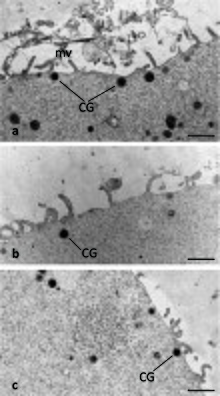
Cortical granules are regulatory secretory organelles found within oocytes and are most associated with polyspermy prevention after fertilization occurs.[1] Cortical granules are found among all mammals, many vertebrates, and some invertebrates.[2] Within the oocyte cortical granules are located along the cortex, the region furthest from the center. Following fertilization, a signaling pathway induces the cortical granules to fuse with the oocyte's cell membrane and release their contents into the oocyte's extracellular matrix.[1] The contents are comprised of proteases, glycosidases, cross-linking enzymes, and structural proteins.[2] This exocytosis of cortical granules is known as the cortical reaction. Evidence suggests the cortical reaction is initiated by both the released Ca(2+) and activated protein kinase C (PKC) following fertilization.[3] Once the cortical granules complete their functions, the oocyte does not replenish them. In mammals, the oocyte's extracellular matrix includes a surrounding layer of perivitelline space, zona pellucida, then cumulus cells. It has been demonstrated experimentally that the released contents of the cortical granules cause cleavage of ZP2, a specific protein found within the zona pellucida contributing to sperm-egg binding. The alteration of the zona pellucida components is known as the zona reaction. Consequently, the zona reaction prevents additional sperm from binding and also impedes the progress of additional sperm already bound. It should also be noted that the cortical reaction does not occur in all mammals, thus suggesting the likelihood of other functional purposes for cortical granules. [1]
Organelle Composition edit
Cortical granules are thought to originate from the oocyte's Golgi apparatus. In some organisms such as in hamsters, the secreted vesicle from the Golgi apparatus may fuse with a secreted vesicle from the rough endoplasmic reticulum to ultimately form a cortical granule.[4]
Once established, the cortical granule maintains a spherical or slightly ovoid shape and is encompassed by a single membrane. Depending on the species, a cortical granule can range from 80-600 µm in diameter. The cortical granules are typically found 2 µm from the oocyte's plasma membrane. However, some cortical granules have been observed to be located further from the plasma membrane[4]
Although the entire cortical granule content has yet to be identified, the following molecules have been found within mammalian cortical granules:[1]
Glycosylated components: Mammalian cortical granules have been shown to contain high levels of carbohydrates. Furthermore, many of these carbohydrates are components of glycosolated molecules such as mannosylated proteins, α-D-acetylgalactosamine, N-acetylglucosamine, N-acetyllactosamine, N-acetylneuraminic acid, D-N-acetylgalactosamine, N-acetylgalactosamine, and N-glycolylneuraminic acid. Certain mannosylated proteins, for instance, are thought to contribute to the cortical granule's envelope structure.[1]
Proteases: Ovastacin (vertebrates?): Cortical granules contain ovastacin, a protease, which cleaves ZP2 following the cortical reaction. Bound to a specific site on ZP2, ovastacin cleaves off ZP2's sperm binding domain, thus preventing any future sperm binding to the zona pellucida.[5][6]
Ovoperoxidase: Ovoperoxidase has been shown to act as a catalyst that cross-links tyrosine residues found within the zone pellucida. This cross-linking contributes to the hardening of the zona pellucida.[1]
Calreticulin:
N-Acetylglucosaminidase
p32:
Peptidylarginine deiminase (PAD/ABL2 antigen/p75):
Types edit
Functions edit
Pre-Fertilzation edit
Fertilization edit
Post-Fertilization edit
Formation edit
Cortical granule formation occurs during the early stages of oocyte growth. More specifically, in the human, monkey, hamster, and rabbit, cortical granules are established once the ovarian follicle is multilayered. In the rat and mouse, cortical granules have been observed earlier in follicle development when the ovarian follicle is only single layered. During the early stages of oocyte growth, the Golgi complex increases in size, proliferates, and produces small vesicles that migrate to the cell's subcortical region. These small vesicles will fuse with one another to form mature cortical granules, which are thus established as separate entities from the Golgi. In mammals, the oocyte continuously produces and translocates cortical granules to the cortex until ovulation occurs. It has been shown in both mammalian and non-mammalian animal models that cortical granule migration depends on cytoskeleton processes, particularly microfilament activity. For mammals, cortical granule migration is considered an indication of oocyte maturity and organelle organization.[1]
Distribution edit
As a result of translocation, cortical granules are evenly distributed throughout the cortex of the oocyte. However, it has been observed in rodents that some cortical granules are rearranged leaving a space amidst the remaining cortical granules. This space is called the cortical granule free domain (CGFD) and has been observed in both the cell's meiotic spindle regions during metaphase I and metaphase II of meiosis. CGFDs have not been observed in feline, equine, bovine, porcine, nor human oocytes. Studies with rodent oocytes suggest that certain cortical granules undergo redistribution and/or exocytosis throughout the meiotic cycle creating the CGFDs. More specifically, evidence includes increased quantities of cortical granules surrounding the CGFDs and a decreased overall quantity of the cell's cortical granules during the meiotic cycle. Additionally, some pre-fertilization cortical granule exocytotic events occur in the cell's cleavage furrow simultaneously with polar body formation.[1]
An assortment of hypotheses exist concerning the biological function of CGFDs and pre-fertilization cortical granule exocytosis. For instance, the formation of the CGFDs may be the oocyte's mechanism for retaining more cortical granules for future use rather than losing them to the polar bodies as they extrude from the cell. Because some cortical granules released are from a region near the meiotic spindles, researchers have also hypothesized that the released cortical granules may modify the oocyte's extracellular matrix so that sperm cannot bind in this region. If sperm were to bind in this region, the paternal DNA, as it decondenses, could possibly disrupt the integrity of the maternal DNA due to its proximity. This blocking of sperm at a specific site is termed local blocking. Considering that rodent oocytes have around 75% less surface area than oocytes of larger mammalian species, sperm binding in this region is more probable thereby possibly necessitating the need for local blocking. Researchers also hypothesize the oocyte releases some cortical granules pre-fertilzation in order to make minor modifications to the oocyte's extracellular matrix so that binding is limited to only sperm capable of binding despite these minor modifications.[1]
Regulation edit
Cortical granule exocytosis is a calcium-dependent event. Upon fertilization, stored calcium within the oocyte is released thereby increasing the calcium level within the cell. This calcium increase occurs as a single wave in echinoderms and as multiple waves in mammals. Cortical granule exocytosis has been shown to occur directly following a calcium wave. For example, in the fertilized sea urchin egg, it has been shown that the cortical granule exocytosis immediately follows the calcium increase after approximately 6 seconds. In mammals, the first calcium wave occurs with 1-4 minutes following fertilization, and cortical granule exocytosis occurs within 5-30 minutes following fertilization. Furthermore, when calcium waves are suppressed experimentally, cortical granule exocytosis and/or alterations in the extracellular matrix did not occur. As demonstrated in unfertilized vertebrate oocytes, cortical granule exocytosis is induced when calcium is artificially increased.[7]
Increased calcium is also thought to activate actin-depolymerizing proteins such as gelsolin and scinderin. In mammals, these actin-depolymerizing proteins serve to disassemble cortical actin thereby allowing space for cortical granule translocation to the plasma membrane.[7]
An oocyte acquires the ability to complete cortical granule exocytosis by the time the oocyte has reached late maturity. More specifically, in mice, for example, the ability to undergo cortical granule exocytosis arises some time between metaphase I and metaphase II of meiosis, which is also 5 hours before ovulation occurs. The oocyte has been shown to obtain maximum proficiency for releasing calcium at this same cell stage, metaphase I and metaphase II, as well, further emphasizing the calcium-dependency of the cortical granule exocytosis event.[7]
History/Discovery edit
References edit
- ^ a b c d e f g h i Liu, Min (17 November 2011). "The biology and dynamics of mammalian cortical granules". Reproductive Biology and Endocrinology. 9 (1). doi:10.1186/1477-7827-9-149. Retrieved 10 March 2015.
{{cite journal}}: CS1 maint: unflagged free DOI (link) - ^ a b Wessel, Gary M.; Brooks, Jacqueline M.; Green, Emma; Haley, Sheila; Voronina, Ekaterina; Wong, Julian; Zaydfudim, Victor; Conner, Sean (2001). "The Biology of Cortical Granules". International Review of Cytology. 209: 117–206. PMID 11580200.
- ^ Sun, Qing-Yuan (1 July 2003). "Cellular and Molecular Mechanisms Leading to Cortical Reaction and Polyspermy Block in Mammalian Eggs". Microscopy Research and Technique. 61 (4): 342–348. PMID 12811739. Retrieved 10 March 2015.
- ^ a b Gulyas, B. J. (1980). "Cortical granules of mammalian eggs". International Review of Cytology. 63: 357–392. PMID 395132. Retrieved 25 March 2015.
- ^ Burkart, Anna D.; Xiong, Bo; Baibakov, Boris; Jiménez-Movilla, Maria; Dean, Jurrien (2 April 2012). "Ovastacin, a cortical granule protease, cleaves ZP2 in the zona pellucida to prevent polyspermy". J Cell Biol. 197 (1): 37–44. doi:10.1083/jcb.201112094. PMID 22472438.
{{cite journal}}:|access-date=requires|url=(help) - ^ Avella, Matteo A.; Xiong, Bo; Dean, Jurrien (May 2013). "The molecular basis of gamete recognition in mice and humans". Molecular Human Reproduction. 19 (5): 279–289. doi:10.1093/molehr/gat004. Retrieved 17 March 2015.
- ^ a b c Abbott, A. L.; Ducibella, T. (1 July 2001). "Calcium and the control of mammalian cortical granule exocytosis". Frontiers in Bioscience. 6 (1): d792-806. doi:10.2741/Abbott. PMID 11438440.
{{cite journal}}:|access-date=requires|url=(help)