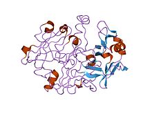
| A1_Propeptide | |||||||||
|---|---|---|---|---|---|---|---|---|---|
 crystal and molecular structures of human progastricsin at 1.62 angstroms resolution | |||||||||
| Identifiers | |||||||||
| Symbol | A1_Propeptide | ||||||||
| Pfam | PF07966 | ||||||||
| InterPro | IPR012848 | ||||||||
| |||||||||
Most eukaryotic endopeptidases (MEROPS peptidase family A1) are synthesised with signal and propeptides. The animal pepsin-like endopeptidase propeptides form a distinct family of propeptides, which contain a conserved motif approximately 30 residues long. In pepsinogen A, the first 11 residues of the mature pepsin sequence are displaced by residues of the propeptide. The propeptide contains two helices that block the active site cleft, in particular the conserved Asp11 residue, in pepsin, hydrogen bonds to a conserved Arg residue in the propeptide. This hydrogen bond stabilises the propeptide conformation and is probably responsible for triggering the conversion of pepsinogen to pepsin under acidic conditions.[1][2]
References edit
- ^ Hartsuck JA, Koelsch G, Remington SJ (May 1992). "The high-resolution crystal structure of porcine pepsinogen". Proteins. 13 (1): 1–25. doi:10.1002/prot.340130102. PMID 1594574.
{{cite journal}}: CS1 maint: date and year (link) CS1 maint: multiple names: authors list (link) - ^ Sielecki AR, Fujinaga M, Read RJ, James MN (June 1991). "Refined structure of porcine pepsinogen at 1.8 A resolution". J. Mol. Biol. 219 (4): 671–692. doi:10.1016/0022-2836(91)90664-r. PMID 2056534.
{{cite journal}}: CS1 maint: date and year (link) CS1 maint: multiple names: authors list (link)