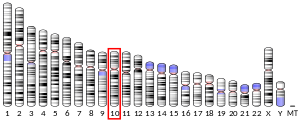
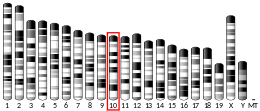
Ten-eleven translocation methylcytosine dioxygenase 1 (TET1) is a member of the TET family of enzymes, in humans it is encoded by the TET1 gene. Its function, regulation, and utilizable pathways remain a matter of current research while it seems to be involved in DNA demethylation and therefore gene regulation.[5][6]
Discovery
editTET1 was first discovered in a 61-year-old patient with a rare variation of t(10;11)(q22;q23) acute myeloid leukemia (AML) as a zinc-finger binding protein (specifically on the CXXC domain) that fuses to the gene MLL.[7] Another study confirmed that this protein was a translocation partner of MLL in an 8-year-old patient with t(10;11)(q22;q23) AML and named the protein Ten-Eleven Translocation 1.[8]
Function
editTET1 catalyzes the conversion of the modified DNA base 5-methylcytosine (5-mC) to 5-hydroxymethylcytosine (5-hmC).[9]
TET1 produces 5-hmC by oxidation of 5-mC in an iron and alpha-ketoglutarate dependent manner.[10] The conversion of 5-mC to 5-hmC has been proposed as the initial step of active DNA demethylation in mammals.[10] Additionally, downgrading TET1 has decreased levels of 5-formylcytosine (5-fC) and 5-carboxylcytosine (5-caC) in both cell cultures and mice.[10]
A site with a 5-hmC base already has increased transcriptional activity, a state termed "functional demethylation". This state is common in post-mitotic neurons.[11]
TET1 may play a role in memory extinction. TET1-knockout mice show markedly impaired memory extinction, despite maintaining normal memory acquisition.[12]
Applications
editTET1 appears to facilitate nuclear reprogramming of somatic cells to iPS cells.[13][14]
The enzyme is also utilized as part of TET-Assisted Bisulfite Sequencing (TAB-seq) to quantify levels of hydroxymethylation in the genome and to distinguish 5-hydroxymethylcytosine (5hmc) from 5-methylcytosine (5mc) at single base resolution. The technique was developed by Chuan He and rectifies the inability of traditional bisulfite sequencing to decipher between the two modified bases. In this technique, TET1 is responsible for the oxidation of 5mc allowing it to be read as thymine following treatment with bisulfite. This is not the case for 5hmc as it is glucosylated in the initial step inhibiting its oxidation by TET1.
Clinical significance
editPatients with schizophrenia or bipolar disorder have shown increased levels of TET1 mRNA and protein expression in the inferior parietal lobule, indicating these diseases may be caused by mistakes in gene expression regulation.[15]
Colon, breast, prostate and liver tumors have significantly reduced levels of TET1 compared to the healthy colon cells and normal epithelial colon cells with downgraded TET1 levels have greater levels of proliferation.[16][17][18][19] Additionally, increasing TET1 expression levels in colon cancer cells decreased cell proliferation in both cell cultures and mice through demethylation of promoters of the WNT signaling pathway.[17]
Breast cancer cell lines with silenced TET1 expression have increased rates of invasion and breast cancers that spread to the lymph nodes are characterized by lower TET1 levels.[20] TET1 levels could be used to detect breast cancer metastasis.[20] A histone deacetylase inhibitor Trichostatin A increased levels of TET1 in breast cancer tissues but was a less effective tumor suppressor in patients with low TET1 expression.[21] Breast cancer patients with high TET1 levels had significantly higher survival probabilities than patients with low TET1 levels.[19]
Degradation of TET1 in hypoxia-induced EMT lung cancer cells led to reduced metastasis rates and cells.[22] Healthy cells transitioning to cancer cells have decreased levels of TET1 but decreasing TET1 expression does not lead to malignancy.[23] Cancer cells using the KRAS pathway had decreased invasive potential after reintroducing TET1, likewise downgrading KRAS increased TET1 levels.[24]
References
edit- ^ a b c GRCh38: Ensembl release 89: ENSG00000138336 – Ensembl, May 2017
- ^ a b c GRCm38: Ensembl release 89: ENSMUSG00000047146 – Ensembl, May 2017
- ^ "Human PubMed Reference:". National Center for Biotechnology Information, U.S. National Library of Medicine.
- ^ "Mouse PubMed Reference:". National Center for Biotechnology Information, U.S. National Library of Medicine.
- ^ "Entrez Gene: Tet methylcytosine dioxygenase 1". Retrieved 2012-07-26.
- ^ Coulter JB, O'Driscoll CM, Bressler JP (October 2013). "Hydroquinone increases 5-hydroxymethylcytosine formation through ten eleven translocation 1 (TET1) 5-methylcytosine dioxygenase". The Journal of Biological Chemistry. 288 (40): 28792–28800. doi:10.1074/jbc.M113.491365. PMC 3789975. PMID 23940045.
- ^ Ono R, Taki T, Taketani T, Taniwaki M, Kobayashi H, Hayashi Y (July 2002). "LCX, leukemia-associated protein with a CXXC domain, is fused to MLL in acute myeloid leukemia with trilineage dysplasia having t(10;11)(q22;q23)". Cancer Research. 62 (14): 4075–4080. PMID 12124344.
- ^ Lorsbach RB, Moore J, Mathew S, Raimondi SC, Mukatira ST, Downing JR (March 2003). "TET1, a member of a novel protein family, is fused to MLL in acute myeloid leukemia containing the t(10;11)(q22;q23)". Leukemia. 17 (3): 637–641. doi:10.1038/sj.leu.2402834. PMID 12646957. S2CID 1202064.
- ^ Tahiliani M, Koh KP, Shen Y, Pastor WA, Bandukwala H, Brudno Y, et al. (May 2009). "Conversion of 5-methylcytosine to 5-hydroxymethylcytosine in mammalian DNA by MLL partner TET1". Science. 324 (5929): 930–935. Bibcode:2009Sci...324..930T. doi:10.1126/science.1170116. PMC 2715015. PMID 19372391.
- ^ a b c Ito S, Shen L, Dai Q, Wu SC, Collins LB, Swenberg JA, et al. (September 2011). "Tet proteins can convert 5-methylcytosine to 5-formylcytosine and 5-carboxylcytosine". Science. 333 (6047): 1300–1303. Bibcode:2011Sci...333.1300I. doi:10.1126/science.1210597. PMC 3495246. PMID 21778364.
- ^ Mellén M, Ayata P, Heintz N (September 2017). "5-hydroxymethylcytosine accumulation in postmitotic neurons results in functional demethylation of expressed genes". Proceedings of the National Academy of Sciences of the United States of America. 114 (37): E7812–E7821. Bibcode:2017PNAS..114E7812M. doi:10.1073/pnas.1708044114. PMC 5604027. PMID 28847947.
- ^ Rudenko A, Dawlaty MM, Seo J, Cheng AW, Meng J, Le T, et al. (September 2013). "Tet1 is critical for neuronal activity-regulated gene expression and memory extinction". Neuron. 79 (6): 1109–1122. doi:10.1016/j.neuron.2013.08.003. PMC 4543319. PMID 24050401.
- ^ Pera MF (December 2013). "Epigenetics, vitamin supplements and cellular reprogramming". Nature Genetics. 45 (12): 1412–1413. doi:10.1038/ng.2834. PMID 24270443. S2CID 11597504.
- ^ Chen J, Gao Y, Huang H, Xu K, Chen X, Jiang Y, et al. (March 2015). "The combination of Tet1 with Oct4 generates high-quality mouse-induced pluripotent stem cells". Stem Cells. 33 (3): 686–698. doi:10.1002/stem.1879. PMID 25331067. S2CID 42714024.
- ^ Dong E, Gavin DP, Chen Y, Davis J (September 2012). "Upregulation of TET1 and downregulation of APOBEC3A and APOBEC3C in the parietal cortex of psychotic patients". Translational Psychiatry. 2 (9): e159. doi:10.1038/tp.2012.86. PMC 3565208. PMID 22948384.
- ^ Yang H, Liu Y, Bai F, Zhang JY, Ma SH, Liu J, et al. (January 2013). "Tumor development is associated with decrease of TET gene expression and 5-methylcytosine hydroxylation". Oncogene. 32 (5): 663–669. doi:10.1038/onc.2012.67. PMC 3897214. PMID 22391558.
- ^ a b Neri F, Dettori D, Incarnato D, Krepelova A, Rapelli S, Maldotti M, et al. (August 2015). "TET1 is a tumour suppressor that inhibits colon cancer growth by derepressing inhibitors of the WNT pathway". Oncogene. 34 (32): 4168–4176. doi:10.1038/onc.2014.356. hdl:2318/150019. PMID 25362856. S2CID 22017396.
- ^ Liu C, Liu L, Chen X, Shen J, Shan J, Xu Y, et al. (2013-05-09). "Decrease of 5-hydroxymethylcytosine is associated with progression of hepatocellular carcinoma through downregulation of TET1". PLOS ONE. 8 (5): e62828. Bibcode:2013PLoSO...862828L. doi:10.1371/journal.pone.0062828. PMC 3650038. PMID 23671639.
- ^ a b Hsu CH, Peng KL, Kang ML, Chen YR, Yang YC, Tsai CH, et al. (September 2012). "TET1 suppresses cancer invasion by activating the tissue inhibitors of metalloproteinases". Cell Reports. 2 (3): 568–579. doi:10.1016/j.celrep.2012.08.030. PMID 22999938.
- ^ a b Sang Y, Cheng C, Tang XF, Zhang MF, Lv XB (2015-01-01). "Hypermethylation of TET1 promoter is a new diagnosic marker for breast cancer metastasis". Asian Pacific Journal of Cancer Prevention. 16 (3): 1197–1200. doi:10.7314/apjcp.2015.16.3.1197. PMID 25735355.
- ^ Lu HG, Zhan W, Yan L, Qin RY, Yan YP, Yang ZJ, et al. (November 2014). "TET1 partially mediates HDAC inhibitor-induced suppression of breast cancer invasion". Molecular Medicine Reports. 10 (5): 2595–2600. doi:10.3892/mmr.2014.2517. PMID 25175940.
- ^ Tsai YP, Chen HF, Chen SY, Cheng WC, Wang HW, Shen ZJ, et al. (December 2014). "TET1 regulates hypoxia-induced epithelial-mesenchymal transition by acting as a co-activator". Genome Biology. 15 (12): 513. doi:10.1186/s13059-014-0513-0. PMC 4253621. PMID 25517638.
- ^ Kudo Y, Tateishi K, Yamamoto K, Yamamoto S, Asaoka Y, Ijichi H, et al. (April 2012). "Loss of 5-hydroxymethylcytosine is accompanied with malignant cellular transformation". Cancer Science. 103 (4): 670–676. doi:10.1111/j.1349-7006.2012.02213.x. PMC 7659252. PMID 22320381. S2CID 5823834.
- ^ Wu BK, Brenner C (December 2014). "Suppression of TET1-dependent DNA demethylation is essential for KRAS-mediated transformation". Cell Reports. 9 (5): 1827–1840. doi:10.1016/j.celrep.2014.10.063. PMC 4268240. PMID 25466250.
Further reading
edit- Abdel-Wahab O, Mullally A, Hedvat C, Garcia-Manero G, Patel J, Wadleigh M, et al. (July 2009). "Genetic characterization of TET1, TET2, and TET3 alterations in myeloid malignancies". Blood. 114 (1): 144–147. doi:10.1182/blood-2009-03-210039. PMC 2710942. PMID 19420352.
- Lorsbach RB, Moore J, Mathew S, Raimondi SC, Mukatira ST, Downing JR (March 2003). "TET1, a member of a novel protein family, is fused to MLL in acute myeloid leukemia containing the t(10;11)(q22;q23)". Leukemia. 17 (3): 637–641. doi:10.1038/sj.leu.2402834. PMID 12646957. S2CID 1202064.
- Morgan AR, Hamilton G, Turic D, Jehu L, Harold D, Abraham R, et al. (September 2008). "Association analysis of 528 intra-genic SNPs in a region of chromosome 10 linked to late onset Alzheimer's disease". American Journal of Medical Genetics. Part B, Neuropsychiatric Genetics. 147B (6): 727–731. doi:10.1002/ajmg.b.30670. PMID 18163421. S2CID 13916214.
- Ono R, Taki T, Taketani T, Taniwaki M, Kobayashi H, Hayashi Y (July 2002). "LCX, leukemia-associated protein with a CXXC domain, is fused to MLL in acute myeloid leukemia with trilineage dysplasia having t(10;11)(q22;q23)". Cancer Research. 62 (14): 4075–4080. PMID 12124344.
- Xu W, Yang H, Liu Y, Yang Y, Wang P, Kim SH, et al. (January 2011). "Oncometabolite 2-hydroxyglutarate is a competitive inhibitor of α-ketoglutarate-dependent dioxygenases". Cancer Cell. 19 (1): 17–30. doi:10.1016/j.ccr.2010.12.014. PMC 3229304. PMID 21251613.
- Guo JU, Su Y, Zhong C, Ming GL, Song H (April 2011). "Hydroxylation of 5-methylcytosine by TET1 promotes active DNA demethylation in the adult brain". Cell. 145 (3): 423–434. doi:10.1016/j.cell.2011.03.022. PMC 3088758. PMID 21496894.
- Frauer C, Rottach A, Meilinger D, Bultmann S, Fellinger K, Hasenöder S, et al. (February 2011). "Different binding properties and function of CXXC zinc finger domains in Dnmt1 and Tet1". PLOS ONE. 6 (2): e16627. Bibcode:2011PLoSO...616627F. doi:10.1371/journal.pone.0016627. PMC 3032784. PMID 21311766.
- Langemeijer SM, Aslanyan MG, Jansen JH (December 2009). "TET proteins in malignant hematopoiesis". Cell Cycle. 8 (24): 4044–4048. doi:10.4161/cc.8.24.10239. PMID 19923888. S2CID 27430810.
- Mohr F, Döhner K, Buske C, Rawat VP (March 2011). "TET genes: new players in DNA demethylation and important determinants for stemness". Experimental Hematology. 39 (3): 272–281. doi:10.1016/j.exphem.2010.12.004. PMID 21168469.


