
Size of this JPG preview of this TIF file: 600 × 600 pixels. Other resolutions: 240 × 240 pixels | 480 × 480 pixels.
Original file (800 × 800 pixels, file size: 1.86 MB, MIME type: image/tiff)
| This is a file from the Wikimedia Commons. Information from its description page there is shown below. Commons is a freely licensed media file repository. You can help. |
Summary
| Description |
English: Oligodendrocytes in rat brain stained with an antibody to myelin basic protein in red and for DNA, showing cell nuclei, in blue. Two oligodendrocyte cell bodies are clearly visible as well as several myelinated axons. These are hollow tubes and so appear as "tramlines" in this confocal image. Image and antibody stain from EnCor Biotechnology Inc. |
| Date | |
| Source | Own work |
| Author | GerryShaw |
Licensing
I, the copyright holder of this work, hereby publish it under the following license:
This file is licensed under the Creative Commons Attribution-Share Alike 4.0 International license.
- You are free:
- to share – to copy, distribute and transmit the work
- to remix – to adapt the work
- Under the following conditions:
- attribution – You must give appropriate credit, provide a link to the license, and indicate if changes were made. You may do so in any reasonable manner, but not in any way that suggests the licensor endorses you or your use.
- share alike – If you remix, transform, or build upon the material, you must distribute your contributions under the same or compatible license as the original.
File history
Click on a date/time to view the file as it appeared at that time.
| Date/Time | Thumbnail | Dimensions | User | Comment | |
|---|---|---|---|---|---|
| current | 01:43, 21 July 2014 | 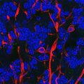 | 800 × 800 (1.86 MB) | GerryShaw | User created page with UploadWizard |
File usage
The following pages on the English Wikipedia use this file (pages on other projects are not listed):
Global file usage
The following other wikis use this file:
- Usage on bs.wikipedia.org
- Usage on hy.wikipedia.org
- Usage on ko.wikipedia.org