
Sir Peter Medawar (1915–1987) described a virus as "a piece of bad news wrapped in a protein coat".[1] With the exception of the bacteriophages, viruses had a well-deserved reputation for being nothing but the cause of diseases and death. The discovery of the abundance of viruses and their overwhelming presence in many ecosystems has led modern virologists to consider them in a new light.[2] They are everywhere. It is estimated that there are 1031 viruses on Earth, most of them are bacteriophages, and most of them are in the oceans.[3] They are the most abundant species on Earth,[4] and they infect all types of organisms, from animals and plants to bacteria and archaea.[5] Since Dmitri Ivanovsky's 1892 article describing a non-bacterial pathogen infecting tobacco plants, and the discovery of the tobacco mosaic virus by Martinus Beijerinck in 1898,[6] about 5,000 viruses have been described in detail,[7] although there are millions of different types.[8] Viruses have existed since since life first appeared on Earth although their origins are unclear.
Viruses are small infectious agents that can replicate only inside living cells. A single virus particle (known as a virion) consists of two or three parts: the genetic material made from either DNA or RNA, long molecules that carry genetic information; a protein coat that protects these genes; and in some cases an envelope of lipids that surrounds the protein coat when they are outside a cell. The average virus is about one one-hundredth the size of the average bacterium. Most viruses are too small to be seen directly with an optical microscope and they are, to all intents and purposes, invisible. It would take 30,000 to 750,000 viruses, side by side, to stretch to 1 centimetre (0.39 in).
Despite their size, the ability of viruses to cause disease and death has changed the course of human history. Their existence has only been known of for about 100 years, but much is now known about them. Although often feared, most viruses exist in peaceful coexistence with their hosts, and they play a key role in maintaining life on Earth.
Etymology edit
The word is from the Latin virus referring to poison and other noxious substances, first used in English in 1392.[9] Virulent, from Latin virulentus (poisonous), dates to 1400.[10] A meaning of "agent that causes infectious disease" is first recorded in 1728,[9] before the discovery of viruses by Dmitri Ivanovsky in 1892. The plural is viruses. The adjective viral dates to 1948.[11] The term virion (plural virions), which dates from 1959,[12] is also used to refer to a single, stable infective viral particle that is released from the cell and is fully capable of infecting other cells of the same type.[13]
Origins and evolution edit
Viruses are ancient. Studies at the molecular level have revealed relationships between viruses infecting organisms from each of the three domains of life, and viral proteins that pre-date the divergence of life and thus the last universal common ancestor.[14] This indicates that viruses emerged early in the evolution of life and existed before modern cells.[15] Viruses are found wherever there is life, [16] but they do not form fossils because they are much smaller than the grains of sedimentary rocks that fossilise plants and animals. The evolution of viruses has had to be traced by other methods. DNA sequencing has been the most powerful, and has provided unexpected insights.[17] Computers are used to measure viral relationships by comparing their DNA or RNA sequences. As would be expected, viruses of the same genus have much of their sequences in common and more distantly related viruses less in common, which is used to draw phylogenetic trees. The mutation rates for many viruses have been measured, allowing estimates to be made of when species of viruses diverged from common ancestors.[18]
There are three main hypotheses that try to explain the origins of viruses:[19][20]
- Regressive hypothesis
- Viruses may have once been small cells that parasitised larger cells. Over time, genes not required by their parasitism were lost. The bacteria rickettsia and chlamydia are living cells that, like viruses, can reproduce only inside host cells. They lend support to this hypothesis, as their dependence on parasitism is likely to have caused the loss of genes that enabled them to survive outside a cell. This is also called the degeneracy hypothesis,[21][22] or reduction hypothesis.[23]
- Cellular origin hypothesis
- Some viruses may have evolved from bits of DNA or RNA that "escaped" from the genes of a larger organism. The escaped DNA could have come from plasmids (pieces of naked DNA that can move between cells) or transposons (molecules of DNA that replicate and move around to different positions within the genes of the cell).[24] Once called "jumping genes", transposons are examples of mobile genetic elements and could be the origin of some viruses. They were discovered in maize by Barbara McClintock in 1950.[25] This is sometimes called the vagrancy hypothesis,[21][26] or the escape hypothesis.[23]
- Coevolution hypothesis
- This is also called the virus-first hypothesis[23] and proposes that viruses may have evolved from complex molecules of protein and nucleic acid at the same time as cells first appeared on Earth and would have been dependent on cellular life for billions of years. Viroids are molecules of RNA that are not classified as viruses because they lack a protein coat. However, they have characteristics that are common to several viruses and are often called subviral agents.[27] Viroids are important pathogens of plants.[28] They do not code for proteins but interact with the host cell and use the host machinery for their replication.[29] The hepatitis delta virus of humans has an RNA genome similar to viroids but has a protein coat derived from hepatitis B virus and cannot produce one of its own. It is, therefore, a defective virus and cannot replicate without the help of hepatitis B virus.[30] In similar manner, the sputnik virophage is dependent on mimivirus, which infects the protozoan Acanthamoeba castellanii.[31] These viruses that are dependent on the presence of other virus species in the host cell are called satellites and may represent evolutionary intermediates of viroids and viruses.[32][33]
There are problems with all of these hypotheses: the regressive hypothesis does not explain why even the smallest of cellular parasites do not resemble viruses in any way. The escape hypothesis does not explain the complex capsids and other structures on virus particles. The virus-first hypothesis contravened the definition of viruses in that they require host cells.[23] Viruses are now recognised as ancient and to have origins that pre-date the divergence of life into the three domains.[34] This discovery has led modern virologists to reconsider and re-evaluate these three classical hypotheses.[34]
The evidence for an ancestral world of RNA cells[35] and computer analysis of viral and host DNA sequences are giving a better understanding of the evolutionary relationships between different viruses and may help identify the ancestors of modern viruses. To date, such analyses have not proved which of these hypotheses is correct.[35] However, it seems unlikely that all currently known viruses have a common ancestor, and viruses have probably arisen numerous times in the past by one or more mechanisms.[36]
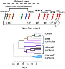
The human genome contains traces of ancient and extinct viruses that once infected hominids and primates, leaving copies of their DNA in the DNA of modern humans; about 31 different families of viruses, called human endogenous retroviruses, have been discovered.[37] Although retroviruses are RNA viruses (which have genes made from RNA not DNA), their reproduction involves a stage where their genes are translated into DNA and inserted into the host cell's DNA.[38] Most of these DNA copies stay in the host cell's genome permanently. A few enter the DNA of the reproductive cells and are passed down through generations of the host's offspring.[39] The last addition to the human genome is estimated to have occurred between 100,000 and 1,000,000 years ago.[37] The discovery of this ancient, once viral, DNA in the human genome has given birth to the science of paleovirology, which although still in its infancy has already provided insights into the co-evolution of humans and viruses and the development of human resistance to them. The study of these "virtual fossils" has become a valuable tool in the study of virus evolution.[37]
Viruses evolve following changes in their DNA (or RNA), some quite rapidly, and the best adapted mutants quickly outnumber their less fit counterparts. In this sense their evolution is Darwinian, just like that of their host organisms.[40] The way viruses reproduce in their host cells makes them particularly susceptible to the genetic changes that help to drive their evolution.[41] The RNA viruses are especially prone to mutations.[42] In host cells there are mechanisms for correcting mistakes when DNA replicates and these kick in whenever cells divide.[42] These important mechanisms prevent potentially lethal mutations from being passed on to offspring. But these mechanisms do not work for RNA and when an RNA virus replicates in its host cell, changes in their genes are occasionally introduced in error, some of which are lethal. One virus particle can produce millions of progeny viruses in just one cycle of replication, therefore the production of a few "dud" viruses is not a problem. Most mutations are "silent" and do not result in any obvious changes to the progeny viruses, but others confer advantages that increase the fitness of the viruses in the environment. These could be changes to the virus particles that disguise them so they are not identified by the cells of the immune system or changes that make antiviral drugs less effective. Both of these changes occur frequently with HIV.[43]

Many viruses (for example, influenza A virus) can "shuffle" their genes with other viruses when two similar strains infect the same cell. This phenomenon is called genetic shift, and is often the cause of new and more virulent strains appearing. Other viruses change more slowly as mutations in their genes gradually accumulate over time, a process known as genetic drift.[45]
Through these mechanisms new viruses are constantly emerging and present a continuing challenge to attempts to control the diseases they cause.[46][47] Most species of viruses are now known to have common ancestors, and although the "virus first" hypothesis has yet to gain full acceptance, there is little doubt that the thousands of species of modern viruses have evolved from less numerous ancient ones.[48] The morbilliviruses, for example, are a group of closely related, but distinct viruses that infect a broad range of animals. The group includes measles virus, which infects humans and primates; canine distemper virus, which infects many animals including dogs, cats, bears, weasels and hyaenas; rinderpest, which infects cattle and buffalo; and other viruses of seals, porpoises and dolphins.[49] Although it not possible to prove which of these rapidly evolving viruses is the earliest, for such a closely related group of viruses to be found in such diverse hosts suggests a possible ancient common ancestor.[50]
Organisms at the edge of life edit
Opinions differ on whether viruses are a form of life, or organic structures that interact with living organisms. They have been described as "organisms at the edge of life",[51] since they resemble organisms in that they possess genes and evolve by natural selection,[52] and reproduce by creating multiple copies of themselves through self-assembly. Although they have genes, they do not have a cellular structure, which is often seen as the basic unit of life. Viruses do not have their own metabolism, and require a host cell to make new products. They therefore cannot naturally reproduce outside a host cell[53] – although bacterial species such as rickettsia and chlamydia are considered living organisms despite the same limitation.[54][55] Accepted forms of life use cell division to reproduce, whereas viruses spontaneously assemble within cells. They differ from autonomous growth of crystals as they inherit genetic mutations while being subject to natural selection. Virus self-assembly within host cells has implications for the study of the origin of life, as it lends further credence to the hypothesis that life could have started as self-assembling organic molecules.[5]
Morphology edit


Viruses display a wide diversity of shapes and sizes, called morphologies. In general, viruses are much smaller than bacteria. Most viruses that have been studied have a diameter between 20 and 300 nanometres. Some filoviruses have a total length of up to 1400 nm; their diameters are only about 80 nm.[56] Most viruses cannot be seen with an optical microscope so scanning and transmission electron microscopes are used to visualise virions.[57] To increase the contrast between viruses and the background, electron-dense "stains" are used. These are solutions of salts of heavy metals, such as tungsten, that scatter the electrons from regions covered with the stain. When virions are coated with stain (positive staining), fine detail is obscured. Negative staining overcomes this problem by staining the background only.[58]

A complete virus particle, known as a virion, consists of nucleic acid surrounded by a protective coat of protein called a capsid. These are formed from identical protein subunits called capsomeres.[59] Viruses can have a lipid "envelope" derived from the host cell membrane. The capsid is made from proteins encoded by the viral genome and its shape serves as the basis for morphological distinction.[60][61] Virally coded protein subunits will self-assemble to form a capsid, in general requiring the presence of the virus genome. Complex viruses code for proteins that assist in the construction of their capsid. Proteins associated with nucleic acid are known as nucleoproteins, and the association of viral capsid proteins with viral nucleic acid is called a nucleocapsid. The capsid and entire virus structure can be mechanically (physically) probed through atomic force microscopy.[62][63] In general, there are four main morphological virus types:


_Virus_PHIL_1878_lores.jpg)
- Helical
- These viruses are composed of a single type of capsomer stacked around a central axis to form a helical structure, which may have a central cavity, or hollow tube. This arrangement results in rod-shaped or filamentous virions: These can be short and highly rigid, or long and very flexible. The genetic material, in general, single-stranded RNA, but ssDNA in some cases, is bound into the protein helix by interactions between the negatively charged nucleic acid and positive charges on the protein. Overall, the length of a helical capsid is related to the length of the nucleic acid contained within it and the diameter is dependent on the size and arrangement of capsomers. The well-studied tobacco mosaic virus is an example of a helical virus.[64]
- Icosahedral
- Most animal viruses are icosahedral or near-spherical with icosahedral symmetry. A regular icosahedron is the optimum way of forming a closed shell from identical sub-units. The minimum number of identical capsomers required is twelve, each composed of five identical sub-units. Many viruses, such as rotavirus, have more than twelve capsomers and appear spherical but they retain this symmetry. Capsomers at the apices are surrounded by five other capsomers and are called pentons. Capsomers on the triangular faces are surrounded by six others and are called hexons.[65] Hexons are in essence flat and pentons, which form the 12 vertices, are curved. The same protein may act as the subunit of both the pentamers and hexamers or they may be composed of different proteins.
- Prolate
- This is an isosahedron elongated along the fivefold axis and is a common arrangement of the heads of bacteriophages. This structure is composed of a cylinder with a cap at either end.[66]
- Envelope
- Some species of virus envelop themselves in a modified form of one of the cell membranes, either the outer membrane surrounding an infected host cell or internal membranes such as nuclear membrane or endoplasmic reticulum, thus gaining an outer lipid bilayer known as a viral envelope. This membrane is studded with proteins coded for by the viral genome and host genome; the lipid membrane itself and any carbohydrates present originate entirely from the host. The influenza virus and HIV use this strategy. Most enveloped viruses are dependent on the envelope for their infectivity.[67]
- Complex
- These viruses possess a capsid that is neither purely helical nor purely icosahedral, and that may possess extra structures such as protein tails or a complex outer wall. Some bacteriophages, such as Enterobacteria phage T4, have a complex structure consisting of an icosahedral head bound to a helical tail, which may have a hexagonal base plate with protruding protein tail fibres. This tail structure acts like a molecular syringe, attaching to the bacterial host and then injecting the viral genome into the cell.[68]

Icosahedral viruses are assigned a triangulation number (T-number) to describe the relationship between the number of protein molecules that form pentagons or hexagons. The T-number idea was originally developed to explain the quasi-symmetry by Caspar and Klug in 1962.[69]

The poxviruses are large, complex viruses that have an unusual morphology. The viral genome is associated with proteins within a central disk structure known as a nucleoid. The nucleoid is surrounded by a membrane and two lateral bodies of unknown function. The virus has an outer envelope with a thick layer of protein studded over its surface. The whole virion is slightly pleiomorphic, ranging from ovoid to brick shape.[70] Mimivirus is the largest characterised virus, with a capsid diameter of 400 nm. Protein filaments measuring 100 nm project from the surface. The capsid appears hexagonal under an electron microscope, therefore the capsid is probably icosahedral.[71] In 2011, researchers discovered a larger virus on ocean floor of the coast of Las Cruces, Chile. Provisionally named Megavirus chilensis, it can be seen with a basic optical microscope. [72]
Some viruses that infect Archaea have complex structures that are unrelated to any other form of virus, with a wide variety of unusual shapes, ranging from spindle-shaped structures, to viruses that resemble hooked rods, teardrops or even bottles. Other archaeal viruses resemble the tailed bacteriophages, and can have multiple tail structures.[73]
Genomes edit
| Property | Parameters |
|---|---|
| Nucleic acid |
|
| Shape |
|
| Strandedness |
|
| Sense |
|
An enormous variety of genomic structures can be seen among viral species; as a group, they contain more structural genomic diversity than plants, animals, archaea, or bacteria. There are millions of different types of viruses,[8] although only about 5,000 of them have been described in detail.[74] A virus has either DNA or RNA genes and is called a DNA virus or a RNA virus, respectively. The vast majority of viruses have RNA genomes. Plant viruses tend to have single-stranded RNA genomes and bacteriophages tend to have double-stranded DNA genomes.[75]
Viral genomes are circular, as in the polyomaviruses, or linear, as in the adenoviruses. The type of nucleic acid is irrelevant to the shape of the genome. Among RNA viruses and certain DNA viruses, the genome is often divided up into separate parts, in which case it is called segmented. For RNA viruses, each segment often codes for only one protein and they are usually found together in one capsid. However, all segments are not required to be in the same virion for the virus to be infectious, as demonstrated by brome mosaic virus and several other plant viruses.[56]
A viral genome, irrespective of nucleic acid type, is almost always either single-stranded or double-stranded. Single-stranded genomes consist of an unpaired nucleic acid, analogous to one-half of a ladder split down the middle. Double-stranded genomes consist of two complementary paired nucleic acids, analogous to a ladder. The virus particles of some virus families, such as those belonging to the Hepadnaviridae, contain a genome that is partially double-stranded and partially single-stranded.[75]
For most viruses with RNA genomes and some with single-stranded DNA genomes, the single strands are said to be either positive-sense (called the plus-strand) or negative-sense (called the minus-strand), depending on whether or not they are complementary to the viral messenger RNA (mRNA). Positive-sense viral RNA is in the same sense as viral mRNA and thus at least a part of it can be immediately translated by the host cell. Negative-sense viral RNA is complementary to mRNA and thus must be converted to positive-sense RNA by an RNA-dependent RNA polymerase before translation. DNA nomenclature for viruses with single-sense genomic ssDNA is similar to RNA nomenclature, in that the coding strand for the viral mRNA is complementary to it (−), and the non-coding strand is a copy of it (+).[75] However, several types of ssDNA and ssRNA viruses have genomes that are ambisense in that transcription can occur off both strands in a double-stranded replicative intermediate. Examples include geminiviruses, which are ssDNA plant viruses and arenaviruses, which are ssRNA viruses of animals.[76]
Genome size varies greatly between species. The smallest viral genomes – the ssDNA circoviruses, family Circoviridae – code for only two proteins and have a genome size of only 2 kilobases; the largest – mimiviruses – have genome sizes of over 1.2 megabases and code for over one thousand proteins.[77] In general, RNA viruses have smaller genome sizes than DNA viruses because of a higher error-rate when replicating, and have a maximum upper size limit.[78] Beyond this limit, errors in the genome when replicating render the virus useless or uncompetitive. To compensate for this, RNA viruses often have segmented genomes – the genome is split into smaller molecules – thus reducing the chance that an error in a single-component genome will incapacitate the entire genome. In contrast, DNA viruses generally have larger genomes because of the high fidelity of their replication enzymes.[79] Single-strand DNA viruses are an exception to this rule, however, as mutation rates for these genomes can approach the extreme of the ssRNA virus case.[80]

Viruses undergo genetic change by several mechanisms. These include a process called genetic drift where individual bases in the DNA or RNA mutate to other bases. Most of these point mutations are "silent" – they do not change the protein that the gene encodes – but others can confer evolutionary advantages such as resistance to antiviral drugs.[81] Antigenic shift occurs when there is a major change in the genome of the virus. This can be a result of recombination or reassortment. When this happens with influenza viruses, pandemics might result.[82] RNA viruses often exist as quasispecies or swarms of viruses of the same species but with slightly different genome nucleoside sequences. Such quasispecies are a prime target for natural selection.[83]
Segmented genomes confer evolutionary advantages; different strains of a virus with a segmented genome can shuffle and combine genes and produce progeny viruses or (offspring) that have unique characteristics. This is called reassortment or viral sex.[84]
Genetic recombination is the process by which a strand of DNA is broken and then joined to the end of a different DNA molecule. This can occur when viruses infect cells simultaneously and studies of viral evolution have shown that recombination has been rampant in the species studied.[85] Recombination is common to both RNA and DNA viruses.[86][87]
Replication cycle edit

Proteins are essential to life. Cells produce new protein molecules from amino acid building blocks based on information coded in DNA. Each type of protein is a specialist that only performs one function, so if a cell needs to do something new, it must make a new protein. Viruses force the cell to make new proteins that the cell does not need, but are needed for the virus to reproduce. Protein synthesis basically consists of two major steps: transcription and translation.
Transcription is the process where information in DNA, called the genetic code, is used to produce RNA copies called messenger RNA (mRNA). These migrate through the cell and carry the code to ribosomes where it is used to make proteins. This is called translation because the protein's amino acid structure is determined by the mRNA's code. Information is hence translated from the language of nucleic acids to the language of amino acids. Some RNA genes of viruses function directly as mRNA without further modification. For this reason, these viruses are called positive-sense RNA viruses.[88] In other RNA viruses, the RNA is a complementary copy of mRNA and these viruses rely on the cell's or their own enzyme to make mRNA. These are called negative-sense RNA viruses. In viruses made from DNA, the method of mRNA production is similar to that of the cell. The species of viruses called retroviruses behave completely differently: they have RNA, but inside the host cell a DNA copy of their RNA is made with the help of the enzyme reverse transcriptase. This DNA is then incorporated into the host's, and copied into mRNA by the cell's normal pathways.[89] Viral populations do not grow through cell division, because they are acellular. Instead, they use the machinery and metabolism of a host cell to produce multiple copies of themselves, and they assemble in the cell.


The life cycle of viruses differs greatly between species but there are six basic stages in the life cycle of viruses:[90]
- Attachment is a specific binding between viral capsid proteins and specific receptors on the host cellular surface. This specificity determines the host range of a virus. For example, HIV infects a limited range of human leucocytes. This is because its surface protein, gp120, specifically interacts with the CD4 molecule – a chemokine receptor – which is most commonly found on the surface of CD4+ T-Cells. This mechanism has evolved to favour those viruses that infect only cells in which they are capable of replication. Attachment to the receptor can induce the viral envelope protein to undergo changes that results in the fusion of viral and cellular membranes, or changes of non-enveloped virus surface proteins that allow the virus to enter.
- Penetration follows attachment: Virions enter the host cell through receptor-mediated endocytosis or membrane fusion. This is often called viral entry. The infection of plant and fungal cells is different from that of animal cells. Plants have a rigid cell wall made of cellulose, and fungi one of chitin, so most viruses can get inside these cells only after trauma to the cell wall.[91] However, nearly all plant viruses (such as tobacco mosaic virus) can also move directly from cell to cell, in the form of single-stranded nucleoprotein complexes, through pores called plasmodesmata.[92] Bacteria, like plants, have strong cell walls that a virus must breach to infect the cell. However, given that bacterial cell walls are much less thick than plant cell walls due to their much smaller size, some viruses have evolved mechanisms that inject their genome into the bacterial cell across the cell wall, while the viral capsid remains outside.[93]
- Uncoating is a process in which the viral capsid is removed: This may be by degradation by viral enzymes or host enzymes or by simple dissociation; the end-result is the releasing of the viral genomic nucleic acid.
- Replication of viruses involves primarily multiplication of the genome. Replication involves synthesis of viral messenger RNA (mRNA) from "early" genes (with exceptions for positive sense RNA viruses), viral protein synthesis, possible assembly of viral proteins, then viral genome replication mediated by early or regulatory protein expression. This may be followed, for complex viruses with larger genomes, by one or more further rounds of mRNA synthesis: "late" gene expression is, in general, of structural or virion proteins.
- Assembly - Following the structure-mediated self-assembly of the virus particles, some modification of the proteins often occurs. In viruses such as HIV, this modification (sometimes called maturation) occurs after the virus has been released from the host cell.[94]
- Release- Viruses can be released from the host cell by lysis, a process that kills the cell by bursting its membrane and cell wall if present: This is a feature of many bacterial and some animal viruses. Some viruses undergo a lysogenic cycle where the viral genome is incorporated by genetic recombination into a specific place in the host's chromosome. The viral genome is then known as a "provirus" or, in the case of bacteriophages a "prophage".[95] Whenever the host divides, the viral genome is also replicated. The viral genome is mostly silent within the host; however, at some point, the provirus or prophage may give rise to active virus, which may lyse the host cells.[96] Enveloped viruses (e.g., HIV) typically are released from the host cell by budding. During this process the virus acquires its envelope, which is a modified piece of the host's plasma or other, internal membrane.[97]
The genetic material within virus particles, and the method by which the material is replicated, varies considerably between different types of viruses.
- DNA viruses
- The genome replication of most DNA viruses takes place in the cell's nucleus. If the cell has the appropriate receptor on its surface, these viruses enter the cell sometimes by direct fusion with the cell membrane (e.g., herpesviruses) or – more usually – by receptor-mediated endocytosis. Most DNA viruses are entirely dependent on the host cell's DNA and RNA synthesising machinery, and RNA processing machinery; however, viruses with larger genomes may encode much of this machinery themselves. In eukaryotes the viral genome must cross the cell's nuclear membrane to access this machinery, while in bacteria it need only enter the cell.[98]
- RNA viruses
- Replication usually takes place in the cytoplasm. RNA viruses can be placed into four different groups depending on their modes of replication. The polarity (whether or not it can be used directly by ribosomes to make proteins) of single-stranded RNA viruses largely determines the replicative mechanism; the other major criterion is whether the genetic material is single-stranded or double-stranded. All RNA viruses use their own RNA replicase enzymes to create copies of their genomes.[99]
- Reverse transcribing viruses
- These have ssRNA (Retroviridae, Metaviridae, Pseudoviridae) or dsDNA (Caulimoviridae, and Hepadnaviridae) in their particles. Reverse transcribing viruses with RNA genomes (retroviruses), use a DNA intermediate to replicate, whereas those with DNA genomes (pararetroviruses) use an RNA intermediate during genome replication. Both types use a reverse transcriptase, or RNA-dependent DNA polymerase enzyme, to carry out the nucleic acid conversion. Retroviruses integrate the DNA produced by reverse transcription into the host genome as a provirus as a part of the replication process; pararetroviruses do not, although integrated genome copies of especially plant pararetroviruses can give rise to infectious virus.[100] They are susceptible to antiviral drugs that inhibit the reverse transcriptase enzyme, e.g. zidovudine and lamivudine. An example of the first type is HIV, which is a retrovirus. Examples of the second type are the Hepadnaviridae, which includes Hepatitis B virus.[101]
Effects on the host cell edit
The range of structural and biochemical effects that viruses have on the host cell is extensive.[102] These are called cytopathic effects.[103] Most virus infections eventually result in the death of the host cell. The causes of death include cell lysis, alterations to the cell's surface membrane and apoptosis.[104] Often cell death is caused by cessation of its normal activities because of suppression by virus-specific proteins, not all of which are components of the virus particle.[105]
Some viruses cause no apparent changes to the infected cell. Cells in which the virus is latent and inactive show few signs of infection and often function normally.[106] This causes persistent infections and the virus is often dormant for many months or years. This is often the case with herpes viruses.[107][108] Some viruses, such as Epstein-Barr virus, can cause cells to proliferate without causing malignancy,[109] while others, such as papillomaviruses, are established causes of cancer.[110]
Host range edit
Viruses are by far the most abundant biological entities on Earth and they outnumber all the others put together.[111] They infect all types of cellular life including animals, plants, bacteria and fungi.[112] However, different types of viruses can infect only a limited range of hosts and many are species-specific. Some, such as smallpox virus for example, can infect only one species – in this case humans,[113] and are said to have a narrow host range. Other viruses, such as rabies virus, can infect different species of mammals and are said to have a broad range.[114] The viruses that infect plants are harmless to animals, and most viruses that infect other animals are harmless to humans.[115] The host range of some bacteriophages is limited to a single strain of bacteria and they can be used to trace the source of outbreaks of infections by a method called phage typing.[116]
Classification edit
Classification seeks to describe the diversity of viruses by naming and grouping them on the basis of similarities. In 1962, André Lwoff, Robert Horne, and Paul Tournier were the first to develop a means of virus classification, based on the Linnaean hierarchical system.[117] This system bases classification on phylum, class, order, family, genus, and species. Viruses were grouped according to their shared properties (not those of their hosts) and the type of nucleic acid forming their genomes.[118] Later the International Committee on Taxonomy of Viruses was formed. However, viruses are not classified on the basis of phylum or class, as their small genome size and high rate of mutation makes it difficult to determine their ancestry beyond Order. As such, the Baltimore Classification is used to supplement the more traditional hierarchy.
ICTV classification edit
The International Committee on Taxonomy of Viruses (ICTV) developed the current classification system and wrote guidelines that put a greater weight on certain virus properties to maintain family uniformity. A unified taxonomy (a universal system for classifying viruses) has been established. The 7th lCTV Report formalised for the first time the concept of the virus species as the lowest taxon (group) in a branching hierarchy of viral taxa.[119] However, at present only a small part of the total diversity of viruses has been studied, with analyses of samples from humans finding that about 20% of the virus sequences recovered have not been seen before, and samples from the environment, such as from seawater and ocean sediments, finding that the large majority of sequences are completely novel.[120]
The general taxonomic structure is as follows:
In the current (2011) ICTV taxonomy, six orders have been established, the Caudovirales, Herpesvirales, Mononegavirales, Nidovirales, Picornavirales and Tymovirales. A seventh order Ligamenvirales has also been proposed. The committee does not formally distinguish between subspecies, strains, and isolates. In total there are 6 orders, 87 families, 19 subfamilies, 349 genera, about 2,284 species and over 3,000 types yet unclassified.[121][122][123]
Baltimore classification edit

The Nobel Prize-winning biologist David Baltimore devised the Baltimore classification system.[124][125] The ICTV classification system is used in conjunction with the Baltimore classification system in modern virus classification.[126][127][128]
The Baltimore classification of viruses is based on the mechanism of mRNA production. Viruses must generate mRNAs from their genomes to produce proteins and replicate themselves, but different mechanisms are used to achieve this in each virus family. Viral genomes may be single-stranded (ss) or double-stranded (ds), RNA or DNA, and may or may not use reverse transcriptase (RT). In addition, ssRNA viruses may be either sense (+) or antisense (−). This classification places viruses into seven groups:
- I: dsDNA viruses (e.g. Adenoviruses, Herpesviruses, Poxviruses)
- II: ssDNA viruses (+)sense DNA (e.g. Parvoviruses)
- III: dsRNA viruses (e.g. Reoviruses)
- IV: (+)ssRNA viruses (+)sense RNA (e.g. Picornaviruses, Togaviruses)
- V: (−)ssRNA viruses (−)sense RNA (e.g. Orthomyxoviruses, Rhabdoviruses)
- VI: ssRNA-RT viruses (+)sense RNA with DNA intermediate in life-cycle (e.g. Retroviruses)
- VII: dsDNA-RT viruses (e.g. Hepadnaviruses)
As an example of viral classification, the chicken pox virus, varicella zoster (VZV), belongs to the order Herpesvirales, family Herpesviridae, subfamily Alphaherpesvirinae, and genus Varicellovirus. VZV is in Group I of the Baltimore Classification because it is a dsDNA virus that does not use reverse transcriptase.
Roles in human diseases edit

Examples of common human diseases caused by viruses include the common cold, influenza, chickenpox and cold sores. Many serious diseases such as ebola, AIDS, avian influenza and SARS are caused by viruses. The relative ability of viruses to cause disease is described in terms of virulence. Other diseases are under investigation as to whether they too have a virus as the causative agent, such as the possible connection between human herpesvirus 6 (HHV6) and neurological diseases such as multiple sclerosis and chronic fatigue syndrome.[131] Viruses have different mechanisms by which they produce disease in an organism, which depends largely on the viral species. Mechanisms at the cellular level primarily include cell lysis, the breaking open and subsequent death of the cell. In multicellular organisms, if enough cells die, the whole organism will start to suffer the effects. Although viruses cause disruption of healthy homeostasis, resulting in disease, they may exist relatively harmlessly within an organism. An example would include the ability of the herpes simplex virus, which causes cold sores, to remain in a dormant state within the human body. This is called latency[132] and is a characteristic of the herpes viruses including Epstein-Barr virus, which causes glandular fever, and varicella zoster virus, which causes chickenpox and shingles. Most people have been infected with at least one of these types of herpes virus.[133] However, these latent viruses might sometimes be beneficial, as the presence of the virus can increase immunity against bacterial pathogens, such as Yersinia pestis.[134] Some viruses can cause lifelong or chronic infections, where the viruses continue to replicate in the body despite the host's defence mechanisms.[135] This is common in hepatitis B virus and hepatitis C virus infections. People chronically infected are known as carriers, as they serve as reservoirs of infectious virus.[136] In populations with a high proportion of carriers, the disease is said to be endemic.[137]
Viruses are an established cause of cancer in humans and other species. Viral cancers occur only in a minority of infected persons (or animals). Cancer viruses come from a range of virus families, including both RNA and DNA viruses, and so there is no single type of "oncovirus" (an obsolete term originally used for acutely transforming retroviruses). The development of cancer is determined by a variety of factors such as host immunity[138] and mutations in the host.[139] Viruses accepted to cause human cancers include some genotypes of human papillomavirus, hepatitis B virus, hepatitis C virus, Epstein-Barr virus, Kaposi's sarcoma-associated herpesvirus and human T-lymphotropic virus. The most recently discovered human cancer virus is a polyomavirus (Merkel cell polyomavirus) that causes most cases of a rare form of skin cancer called Merkel cell carcinoma.[140] Hepatitis viruses can develop into a chronic viral infection that leads to liver cancer.[141][142] Infection by human T-lymphotropic virus can lead to tropical spastic paraparesis and adult T-cell leukemia.[143] Human papillomaviruses are an established cause of cancers of cervix, skin, anus, and penis.[144] Within the Herpesviridae, Kaposi's sarcoma-associated herpesvirus causes Kaposi's sarcoma and body cavity lymphoma, and Epstein–Barr virus causes Burkitt's lymphoma, Hodgkin’s lymphoma, B lymphoproliferative disorder, and nasopharyngeal carcinoma.[145] Merkel cell polyomavirus, which is closely related to the simian SV40 virus, and mouse polyomaviruses have been used as animal models for cancer viruses for over 50 years.[146]
Diagnosis edit

For many virus infections, diagnosis can be made from the signs and symptoms alone. Some infections are more difficult to diagnose and laboratory tests are required. In humans and other animals, blood tests are often used because when the adaptive immune system of a vertebrate encounters a virus, it produces specific antibodies which bind to the virus and render it non-infectious. Two types of antibodies are important. The first called IgM is highly effective at combating viruses but is only produced by the cells of the immune system for a few weeks. The second, called, IgG is produced indefinitely. The presence of IgM in the blood of the host is used to test for acute infection, whereas IgG indicates an infection sometime in the past.[147] Both types of antibodies are measured when tests for immunity are carried out.[148] Other tests include those that allow the detection of the virus's DNA (or RNA) in samples of blood and other bodily fluids, and those that can react with with parts of the viruses called antigens that can be found in infected fluids and cells.[149]
Epidemiology edit

Viral epidemiology is the branch of medical science that deals with the transmission and control of virus infections in humans. Transmission of viruses can be vertical, that is from mother to child, or horizontal, which means from person to person. Examples of vertical transmission include hepatitis B virus and HIV where the baby is born already infected with the virus.[150] Another, rarer, example is the varicella zoster virus, which, although causing relatively mild infections in humans, can be fatal to the foetus and new-born baby.[151]
Horizontal transmission is the most common mechanism of spread of viruses in populations. Transmission can occur when: body fluids are exchanged during sexual activity, e.g., HIV; blood is exchanged by contaminated transfusion or needle sharing, e.g., hepatitis C; exchange of saliva by mouth, e.g., Epstein-Barr virus; contaminated food or water is ingested, e.g., norovirus; aerosols containing virions are inhaled, e.g., influenza virus; and insect vectors such as mosquitoes penetrate the skin of a host, e.g., dengue. The rate or speed of transmission of viral infections depends on factors that include population density, the number of susceptible individuals, (i.e., those not immune),[152] the quality of healthcare and the weather.[153]
Epidemiology is used to break the chain of infection in populations during outbreaks of viral diseases.[154] Control measures are used that are based on knowledge of how the virus is transmitted. It is important to find the source, or sources, of the outbreak and to identify the virus. Once the virus has been identified, the chain of transmission can sometimes be broken by vaccines. When vaccines are not available sanitation and disinfection can be effective. Often infected people are isolated from the rest of the community and those that have been exposed to the virus placed in quarantine.[155] To control the outbreak of foot-and-mouth disease in cattle in Britain in 2001, thousands of cattle were slaughtered.[156] Most viral infections of humans and other animals have incubation periods during which the infection causes no signs or symptoms.[157] Incubation periods for viral diseases range from a few days to weeks but are known for most infections.[158] Somewhat overlapping, but mainly following the incubation period, there is a period of communicability; a time when an infected individual or animal is contagious and can infect another person or animal.[158] This too is known for many viral infections and knowledge the length of both periods is important in the control of outbreaks.[159] When outbreaks cause an unusually high proportion of cases in a population, community or region they are called epidemics. If outbreaks spread worldwide they are called pandemics.[160]
Host defence mechanisms edit
The body's first line of defence against viruses is the innate immune system. This comprises cells and other mechanisms that defend the host from infection in a non-specific manner. This means that the cells of the innate system recognise, and respond to, pathogens in a generic way, but, unlike the adaptive immune system, it does not confer long-lasting or protective immunity to the host.[161]
RNA interference is an important innate defence against viruses.[162] Many viruses have a replication strategy that involves double-stranded RNA (dsRNA). When such a virus infects a cell, it releases its RNA molecule or molecules, which immediately bind to a protein complex called dicer that cuts the RNA into smaller pieces. A biochemical pathway called the RISC complex is activated, which degrades the viral mRNA and the cell survives the infection. Rotaviruses avoid this mechanism by not uncoating fully inside the cell and by releasing newly produced mRNA through pores in the particle's inner capsid. The genomic dsRNA remains protected inside the core of the virion.[163][164]
When the adaptive immune system of a vertebrate encounters a virus, it produces specific antibodies that bind to the virus and render it non-infectious. This is called humoral immunity. Two types of antibodies are important. The first, called IgM, is highly effective at neutralizing viruses but is produced by the cells of the immune system only for a few weeks. The second, called IgG, is produced indefinitely. The presence of IgM in the blood of the host is used to test for acute infection, whereas IgG indicates an infection sometime in the past.[165] IgG antibody is measured when tests for immunity are carried out.[166]

A second defence of vertebrates against viruses is called cell-mediated immunity and involves immune cells known as T cells. The body's cells constantly display short fragments of their proteins on the cell's surface, and, if a T cell recognises a suspicious viral fragment there, the host cell is destroyed by killer T cells and the virus-specific T-cells proliferate. Cells such as the macrophage are specialists at this antigen presentation.[167] The production of interferon is an important host defence mechanism. This is a hormone produced by the body when viruses are present. Its role in immunity is complex; it eventually stops the viruses from reproducing by killing the infected cell and its close neighbours.[168]
Not all virus infections produce a protective immune response in this way. HIV evades the immune system by constantly changing the amino acid sequence of the proteins on the surface of the virion. These persistent viruses evade immune control by sequestration, blockade of antigen presentation, cytokine resistance, evasion of natural killer cell activities, escape from apoptosis, and antigenic shift.[169] Other viruses, called neurotropic viruses, are disseminated by neural spread where the immune system may be unable to reach them.
Many virus infections are spread from person to person and immunity in a proportion of the population can protect susceptible individuals because the chain of transmission is broken. This is known as herd immunity. The reproductive rate – the number of further cases caused by an infected person - is known for many virus infections. If this rate is reduced to less than one, the infection will stop spreading. And if humans are the only host for the virus, the disease can be eradicated.[170]
| Disease | Transmission | R0 | Herd immunity threshold |
|---|---|---|---|
| Measles | Airborne | 12–18 | 83–94% |
| Mumps | Airborne droplet | 4–7 | 75–86% |
| Polio | Fecal-oral route | 5–7 | 80–86% |
| Rubella | Airborne droplet | 5–7 | 80–85% |
| Smallpox | Social contact | 6–7 | 83–85% |
| ^ - R0 is the basic reproduction number, or the average number of secondary infectious cases that are produced by a single index case in completely susceptible population. | |||
Prevention and treatment edit
Because viruses use vital metabolic pathways within host cells to replicate, they are difficult to eliminate without using drugs that cause toxic effects to host cells in general. The most effective medical approaches to viral diseases are vaccinations to provide immunity to infection, and antiviral drugs that selectively interfere with viral replication.[172][173]
Vaccines edit
Vaccination is a cheap and effective way of preventing infections by viruses. Vaccines were used to prevent viral infections long before the discovery of the actual viruses. Their use has resulted in a dramatic decline in morbidity (illness) and mortality (death) associated with viral infections such as polio, measles, mumps and rubella.[174] Smallpox infections have been eradicated.[175] Vaccines are available to prevent over thirteen viral infections of humans,[176] and more are used to prevent viral infections of animals.[177] Vaccines can consist of live-attenuated or killed viruses, or viral proteins (antigens).[178] Live vaccines contain weakened forms of the virus, which do not cause the disease but, nonetheless, confer immunity. Such viruses are called attenuated. Live vaccines can be dangerous when given to people with a weak immunity, (who are described as immunocompromised), because in these people, the weakened virus can cause the original disease.[179] Biotechnology and genetic engineering techniques are used to produce subunit vaccines. These vaccines use only the capsid proteins of the virus. Hepatitis B vaccine is an example of this type of vaccine.[180] Subunit vaccines are safe for immunocompromised patients because they cannot cause the disease.[181] The yellow fever virus vaccine, a live-attenuated strain called 17D, is probably the safest and most effective vaccine ever generated.[182]

Antiviral drugs edit


Antiviral drugs are often nucleoside analogues, (fake DNA building-blocks), which viruses mistakenly incorporate into their genomes during replication. The life-cycle of the virus is then halted because the newly synthesised DNA is inactive. This is because these analogues lack the hydroxyl groups, which, along with phosphorus atoms, link together to form the strong "backbone" of the DNA molecule. This is called DNA chain termination.[183] Examples of nucleoside analogues are aciclovir for Herpes simplex virus infections and lamivudine for HIV and Hepatitis B virus infections. Aciclovir is one of the oldest and most frequently prescribed antiviral drugs.[184] Other antiviral drugs in use target different stages of the viral life cycle. HIV is dependent on a proteolytic enzyme called the HIV-1 protease for it to become fully infectious. There is a large class of drugs called protease inhibitors that inactivate this enzyme.[185]
Hepatitis C is caused by an RNA virus. In 80% of people infected, the disease is chronic, and without treatment, they are infected for the remainder of their lives. However, there is now an effective treatment that uses the nucleoside analogue drug ribavirin combined with interferon.[186] The treatment of chronic carriers of the hepatitis B virus by using a similar strategy using lamivudine has been developed.[187]
Infection in other species edit
Viruses infect all cellular life and, although viruses occur universally, each cellular species has its own specific range that often infect only that species.[188] Some viruses, called satellites, can replicate only within cells that have already been infected by another virus.[31] Viruses are important pathogens of livestock. Diseases such as foot-and-mouth disease and bluetongue are caused by viruses.[189] Companion animals such as cats, dogs, and horses, if not vaccinated, are susceptible to serious viral infections. Canine parvovirus is caused by a small DNA virus and infections are often fatal in pups.[190] Like all invertebrates, the honey bee is susceptible to many viral infections.[191] However, most viruses co-exist harmlessly in their host and cause no signs or symptoms of disease.[192]
Plants edit

There are many types of plant virus, but often they cause only a loss of yield, and it is not economically viable to try to control them. Plant viruses are often spread from plant to plant by organisms, known as vectors. These are normally insects, but some fungi, nematode worms, and single-celled organisms have been shown to be vectors. When control of plant virus infections is considered economical, for perennial fruits, for example, efforts are concentrated on killing the vectors and removing alternate hosts such as weeds.[193] Plant viruses cannot infect humans and other animals because they can reproduce only in living plant cells.[194]
Plants have elaborate and effective defence mechanisms against viruses. One of the most effective is the presence of so-called resistance (R) genes. Each R gene confers resistance to a particular virus by triggering localised areas of cell death around the infected cell, which can often be seen with the unaided eye as large spots. This stops the infection from spreading.[195] RNA interference is also an effective defence in plants.[196] When they are infected, plants often produce natural disinfectants that kill viruses, such as salicylic acid, nitric oxide, and reactive oxygen molecules.[197]
Plant virus particles or virus-like particles (VLPs) have applications in both biotechnology and nanotechnology. The capsids of most plant viruses are simple and robust structures and can be produced in large quantities either by the infection of plants or by expression in a variety of heterologous systems. Plant virus particles can be modified genetically and chemically to encapsulate foreign material and can be incorporated into supramolecular structures for use in biotechnology.[198]
Bacteria edit

Bacteriophages are a common and diverse group of viruses and are the most abundant form of biological entity in aquatic environments – there are up to ten times more of these viruses in the oceans than there are bacteria,[199] reaching levels of 250,000,000 bacteriophages per millilitre of seawater.[200] These viruses infect specific bacteria by binding to surface receptor molecules and then entering the cell. Within a short amount of time, in some cases just minutes, bacterial polymerase starts translating viral mRNA into protein. These proteins go on to become either new virions within the cell, helper proteins, which help assembly of new virions, or proteins involved in cell lysis. Viral enzymes aid in the breakdown of the cell membrane, and, in the case of the T4 phage, in just over twenty minutes after injection over three hundred phages could be released.[201]
The major way bacteria defend themselves from bacteriophages is by producing enzymes that destroy foreign DNA. These enzymes, called restriction endonucleases, cut up the viral DNA that bacteriophages inject into bacterial cells.[202] Bacteria also contain a system that uses CRISPR sequences to retain fragments of the genomes of viruses that the bacteria have come into contact with in the past, which allows them to block the virus's replication through a form of RNA interference.[203][204] This genetic system provides bacteria with acquired immunity to infection.
Archaea edit
Some viruses replicate within archaea: these are double-stranded DNA viruses with unusual and sometimes unique shapes.[205][73] These viruses have been studied in most detail in the thermophilic archaea, particularly the orders Sulfolobales and Thermoproteales.[206] Defences against these viruses may involve RNA interference from repetitive DNA sequences within archaean genomes that are related to the genes of the viruses.[207][208]
Role in aquatic ecosystems edit
.jpg)
A teaspoon of seawater contains about one million viruses.[209] They are essential to the regulation of saltwater and freshwater ecosystems.[210] Most of these viruses are bacteriophages, which are harmless to plants and animals. They infect and destroy the bacteria in aquatic microbial communities, comprising the most important mechanism of recycling carbon in the marine environment. The organic molecules released from the bacterial cells by the viruses stimulate fresh bacterial and algal growth.[211]
Microorganisms constitute more than 90% of the biomass in the sea. It is estimated that viruses kill approximately 20% of this biomass each day and that there are 15 times as many viruses in the oceans as there are bacteria and archaea. Viruses are the main agents responsible for the rapid destruction of harmful algal blooms,[212] which often kill other marine life.[213] The number of viruses in the oceans decreases further offshore and deeper into the water, where there are fewer host organisms.[214] The effects of marine viruses are far-reaching; by increasing the amount of photosynthesis in the oceans, viruses are indirectly responsible for reducing the amount of carbon dioxide in the atmosphere by approximately 3 gigatonnes of carbon per year.[214] Viruses are efficient gene donators and it has been estimated that 10 percent of the world's photosynthesis is driven by genes that originated in marine bacteriophges.[215] The oceans are a vast untapped reservoir viral diversity and the important role that viruses play in marine ecology has become a subject of fruitful research.[216]
Like any organism, marine mammals are susceptible to viral infections. In 1988 and 2002, thousands of harbor seals were killed in Europe by phocine distemper virus.[217] Many other viruses, including caliciviruses, herpesviruses, adenoviruses and parvoviruses, circulate in marine mammal populations.[214]
Role in evolution edit
Viruses are an important natural means of transferring genes between different species, which increases genetic diversity and drives evolution.[218] It is thought that viruses played a central role in the early evolution, before the diversification of bacteria, archaea and eukaryotes and at the time of the last universal common ancestor of life on Earth.[219] Viruses are still one of the largest reservoirs of unexplored genetic diversity on Earth.[214][220]
Extraterrestrials edit

In 1979, the famous physicist Sir Fred Hoyle and N.C Wickramasinghe, first proposed their theory of microbial panspermia in that virus pandemics originate in outer space.[221] Although some hospital consultants in charge of infection control might secretly think this is true, the theory was soon discredited by virologists who used more proven epidemiology.[222] But for life, to exist – "as we know it"[223] – it needs water, energy and a rich source of carbon and some trace elements.[224] So, given the size of the universe, there seems little doubt that life as we know it exists elsewhere.[225] When, in 1969, astronauts Armstrong, Aldrin and Collins returned from the moon, they were quarantined for 21 days in case they had been infected with lunar microbes. But these fears were unjustified and subsequent lunar explorers were not subjected to the indignity.[226] The possibility of the existence of viruses on other worlds cannot be dismissed but it is unlikely that these would pose a threat to life on earth because of their co-evolution with their hosts and subsequent host specificity. Given that the earth-bound tomato bushy stunt virus has such a narrow host range, it is hard to conceive that extraterrestrial viruses might have a specificity broad enough to enable them to infect life on Earth.[227]
Viruses in history edit
Prehistory edit

Over the past 50,000–100,000 years as modern humans dispersed throughout the world new infectious diseases, including those caused by viruses, emerged.[228] Smallpox, which was one of the most lethal and devastating viral infections, might have emerged first among agricultural communities in Africa in about 10,000 BC.[229] The virus, which only infected humans, probably descended from the poxviruses of rodents.[230] Humans probably came into contact with these rodents, and some people became infected by the viruses they carried.
When viruses cross this so called "species barrier" their effects can be severe,[231] and humans may have had little natural resistance. Contemporary humans lived in small communities, and those who succumbed to infection either died or developed immunity. In humans, this acquired immunity is only passed down to offspring temporarily through breast milk and the antibodies, which cross the placenta from the mother's blood to the unborn child's. Therefore, sporadic outbreaks probably occurred in each generation. In about 9000 BC, when many people began to settle on the fertile flood plains of the River Nile, the population became dense enough for the virus to maintain a constant presence thanks to the high concentration of susceptible people.[232]
The Neolithic age, which began in the Middle East in about 9500 BC, was a time when humans became farmers. This agricultural revolution embraced the development of monoculture and presented an opportunity for the rapid spread of several species of plant viruses.[233] The divergence and spread of sobemoviruses – southern bean mosaic virus – date from this time[234] and the spread of the potyviruses of potatoes, and other fruits and vegetables, began about 6,600 years ago.[233]
About 10,000 years ago the humans who inhabited the lands around the Mediterranean basin began to domesticate wild animals. Pigs, cattle, goats, sheep, horses, camels, cats and dogs were all kept and bred in captivity.[235] These animals would have brought their viruses with them. The transmission of viruses from animals to humans can occur, but such zoonotic infections are rare and subsequent human-to-human transmission is even rarer, although there are notable exceptions such as influenza. Most viruses are species specific and would have posed no threat to humans. But as humans became more dependent on domesticated animals, any outbreaks of disease among their livestock – in animals these are called epizootics – could have devastating consequences.
Other, more ancient, viruses are less of a threat. Humans have lived with herpes virus infections since humans first came into being. The virus passed to us from other mammals more than 80 million years ago.[236] Humans have developed a tolerance to these viruses, and most are infected with at least one species of them without being aware of it. Records of these milder virus infections are understandably rare. But there is no reason to doubt that early hominids suffered from colds, 'flu and diarrhoea caused by viruses just as humans do today. It is the younger viruses that cause epidemics and pandemics – and it is those that history records.[236]
Antiquity edit

Among the earliest records of a viral infection is an Egyptian stele thought to depict an Egyptian priest from the 18th Dynasty (1580–1350 BC) with a foot drop deformity characteristic of a poliovirus infection.[237] The mummy of Siptah – a ruler during the 19th Dynasty – shows signs of poliomyelitis, and that of Ramesses V and some other Egyptian mummies buried over 3000 years ago show evidence of smallpox.[238] There was an epidemic of smallpox in Athens in 430 BC. A quarter of the Athenian army died from the infection along with many of the city's civilians.[239]

Measles is an old disease, but it was not until the 10th century that the Persian physician Muhammad ibn Zakariya al-Razi (865–925) (known as "Rhazes") first identified it.[240] Rhazes used the Arabic name "hasbah" for measles, but it has had many other names including "rubeola" from the Latin word rubeus, which means red, and "morbilli", which means "small plague".[241] The close similarities between measles virus, canine distemper virus, and rinderpest virus have given rise to speculation that measles was first transmitted to humans from domesticated dogs or cattle.[242] The measles virus appears to have fully diverged from the then-widespread rinderpest virus by the 11th and 12th centuries.[243] The earliest likely origin is within the seventh century: for this earlier origin there is some linguistic evidence.[244][245]
One measles infection confers lifelong immunity, therefore the virus requires a high population density to become endemic and probably did not occur in the Neolithic.[240] (In more recent times, measles has become extinct on remote islands with populations of less than 500,000 people.[242]) Following the emergence of the virus in the Middle East it reached India by 2500 BC.[246] Measles was so common in children at the time it was not recognised as a disease. In Egyptian hieroglyphs it was described as a normal stage of human development.[247] The Americas and Australia remained free of measles and smallpox until the arrival of European colonists between the 15th and 18th centuries.[228]
One of the earliest descriptions of a virus-infected plant can be found in a poem written by the Japanese Empress Kōken (718–770), in which she describes a plant in summer with yellowing leaves. The plant, later identified as Eupatorium lindleyanum, is often infected with Tomato yellow leaf curl virus.[248]
Middle Ages edit

The Middle Ages witnessed many plagues and pestilences. The rapidly growing population of Europe and the rising concentrations of people in its towns and cities became a fertile ground for many infectious and contagious diseases, of which the Black Death – a bacterial infection – is probably the most notorious.[249] Except for smallpox and influenza, documented outbreaks of infections now known to be caused by viruses are rare. Rabies, a disease that had been recognised for over 4000 years,[250] was rife in Europe, and continued to be until the development of a vaccine by Louis Pasteur in 1886.[251] The average life expectancy during the Middle Ages was 35 years and 60% of children died before the age of 16, many of them during their first 6 years of life. Among the plethora of diseases common at the time were influenza, measles, and smallpox.[252]
Measles was endemic throughout the highly populated countries of Europe, North Africa, and the Middle East. In England the disease, then called "mezils", was first described in the 13th century, and was probably one of the 49 plagues that occurred between 526 and 1087.[246] Rinderpest has been recognised since Roman times.[253] The disease, which originated in Asia, was first brought to Europe by the invading Huns in 370. Later invasions of Mongols, led by Genghis Khan and his army, started pandemics in Europe in 1222, 1233 and 1238. The infection subsequently reached England following the importation of cattle from Europe.[254] At the time rinderpest was a devastating disease with a mortality rate of 80–90%. The resulting loss of cattle cased famine and starvation.[254]
Early modern period edit

Many paintings can be found in the museums of Europe depicting tulips with attractive coloured stripes. Most, such as the still life studies of Johannes Bosschaert, were painted during the 17th century. These flowers were particularly popular and became sought after by those who could afford them. It was not known at the time that these attractive – and very expensive – stripes were caused by a virus accidentally transferred by humans to tulips from jasmine.[255]
Until the Irish Great Famine of 1845–1852, the commonest cause of disease in potatoes was not the mould that causes blight but a virus. The disease, called "curl", is caused by Potato leafroll virus, and it was widespread in England in the 1770s, where it destroyed 75% of the potato crop. At the time, the Irish potato crop remained relatively unscathed.[256]

In 1546 Girolamo Fracastoro (1478–1553) wrote a classic description of measles. He thought the disease was caused by "seeds" (seminaria) that were spread from person to person. An epidemic hit London in 1670, recorded by Thomas Sydenham (1624–1689), who thought it was caused by toxic vapours emanating from the earth.[246] Sydenham had been a soldier and fought for the Parliamentarians during the English Civil War. As a physician, he was a skilled observer and kept meticulous records.[257]
A short time after Henry Tudor's victory at the Battle of Bosworth on 22 August 1485, his army suddenly went down with "the English sweat", which contemporary observers described as something new.[258] It was probably influenza, although we cannot be sure,[259] because records from the time when medicine was not a science can be unreliable.[260] The language used to describe diseases in these sources is often vague and colloquial. We read of "plagues of mice", "changes in the direction of the wind" and the "influence of comets". This makes retrospective diagnosis difficult. As medicine became a science, the descriptions of disease became less vague and records became more reliable.
References to influenza infections date from the late 15th and early 16th centuries,[261] but infections almost certainly occurred long before then.[262] The first that was reliably recorded began in Malta in July 1580, and swept across Europe, Africa, and Asia.[263] More than a century passed before the three pandemics of the 18th century; the one that took place during 1781–2 was probably the most devastating in history.[264] The pandemic began in November 1781 in the East Indies and reached Moscow in December. In February 1782 it hit St. Petersburg and by May it had reached Denmark.[265] Within six weeks, three-quarters of the British population were infected and the pandemic soon spread to the Americas.[266]

Along with measles and influenza, smallpox was taken to the Americas by the Spanish.[267] Smallpox was endemic in Spain, having been introduced by the Moors from Africa.[268] In 1519, an epidemic of smallpox broke out in the Aztec capital Tenochtitlan, which was started by the army of Pánfilo de Narváez (1478–1528), who followed Hernán Cortés (1495–1587) from Cuba, and had an African slave suffering from smallpox aboard his ship.[268] When the Spanish finally entered the capital in the summer of 1521, they saw it strewn with the bodies of smallpox victims.[269] The epidemic, and those that followed during 1545–1548 and 1576–1581, eventually killed more than half of the native population.[270] Most of the Spanish were immune and with his army of fewer than 900 men, it would not have been possible for Cortés to defeat the Aztecs and conquer Mexico without the help of smallpox.[271]
History repeated itself, and many Native American populations were devastated by the spread of smallpox, measles, and influenza.[228] There were, on average, fourteen epidemics of smallpox in the 18th century and by 1733, the disease had affected all Six Nations. In 1738, half the population of Cherokee died from the disease. In Pennsylvania, in 1763, Europeans intentionally exposed Native Americans to smallpox.[272] It is not known exactly how many Native Americans were killed, but the damage done by this disease significantly aided European attempts to displace and conquer the native population.[273]

By the 18th century, smallpox was endemic in Europe. There were five epidemics in London between 1719 and 1746, and large outbreaks occurred in other major European cities. By the end of the century about 400,000 Europeans were dying from the disease each year.[274] It reached South Africa in 1713, having been carried by ships from India, and decimated the population of Hottentots in 1713 and 1755.[275] In 1789 the disease struck Australia,[274] and in the 19th century, smallpox became the single major cause of death of the Australian Aborigines.[276]
Yellow fever is an often lethal disease caused by a flavivirus. The virus is transmitted to humans by mosquitoes (Aedes aeypti) and first appeared over 3,000 years ago.[277] In 1647, the first recorded epidemic occurred on Barbados and was called "Barbados distemper" by John Winthrop (1588–1649), who was the governor of the island at the time. He passed quarantine laws to protect the people – the first ever such laws in North America.[278] Further epidemics of the disease occurred in the North Americas in the 17th, 18th, and 19th centuries.[279]
The first known cases of dengue fever occurred in Indonesia and Egypt in 1779 and trade ships brought the disease to the US, where an epidemic occurred in Philadelphia in 1780.[280] In the 19th century there were frequent dengue epidemics throughout the Americas, and by 1897 almost the whole of the southern US was affected.[280]
Discovery of vaccination edit

Lady Mary Wortley Montagu (1689–1762) was an aristocrat, a writer and the wife of a Member of Parliament. In 1716, her husband, Edward Wortley Montagu was appointed British Ambassador in Istanbul. She followed him there and two weeks after her arrival discovered the local practice of protection against smallpox by variolation – the injection of pus from smallpox victims into the skin.[232] Her younger brother had died of smallpox, and she too had had the disease but survived. Determined to spare her five-year-old son Edward from similar suffering, she ordered the embassy surgeon, Charles Maitland to variolate him. On her return to London, she asked Maitland to variolate her four-year-old daughter in the presence of the king's physicians.[281] Later, Montagu persuaded the Prince and Princess of Wales to sponsor a public demonstration of the procedure. Six prisoners who had been condemned to death and were awaiting execution at Newgate Prison were offered a full pardon for serving as the subjects of the public experiment. They accepted, and in 1721 were variolated. All the prisoners recovered from the procedure and to test its protective effect one of them, a nineteen-year-old woman, was ordered to sleep in the same bed as a ten-year-old smallpox victim for six weeks. She did not contract the disease.[282] The experiment was repeated on six orphan children who survived the ordeal and by 1722, even King George I's grandchildren had been inoculated. But the practice was not entirely safe and two percent died as a result.[283] Although the number of lives saved far outweighed those lost, the practice was not widely adopted.[284]

At the time nothing was known about viruses or immunity and no one knew how variolation worked.[285]
Edward Jenner (1749–1823), a British rural physician, was variolated as a boy. He had suffered greatly from the ordeal but survived fully protected from smallpox. Jenner knew of a local belief that dairy workers who had contracted a relatively mild infection called cowpox were immune to smallpox. He decided to test the "theory" (though he was probably not the first to do). On 14 May 1796 he selected "a healthy boy, about eight years old for the purpose of inoculation for the Cow Pox".[286] The boy was James Phipps (1788–1853) who survived the experiment and suffered only a mild fever. On 1 July 1796, Jenner took some "smallpox matter" (probably infected pus) and repeatedly inoculated Phipp's arms with it. Phipps survived, and was subsequently inoculated with smallpox more than 20 times without succumbing to the disease. Vaccination – the word is derived from the Latin vacca meaning "cow" – had been invented.[287]
Louis Pasteur and rabies edit

Rabies is an often fatal disease caused by the infection of mammals with rabies virus. Today it is mainly a disease that affects wild mammals such as foxes and bats, but it is one of the oldest known virus diseases: rabies is a Sanskrit word (rabhas) that dates from 3000 BC,[251] which means "madness" or "rage"[247] and the disease has been known for over 4000 years.[250] The ancient Greeks called it "lyssa" or "lytta" also meaning madness.[250] Although not a cause of epidemics the infection was greatly feared because of its horrendous symptoms, which include insanity, hydrophobia, and death.[250] The disease had been known since antiquity, and references to rabies can be found in the Laws of Eshnunna, which date from 2300 BC. Aristotle (384–322 BC) wrote one of earliest undisputed descriptions of the disease and how it was passed to humans. Celsus in the first century AD first recorded the symptom called hydrophobia and suggested the saliva of infected animals and humans contained a slime or poison – to describe this he used the word "virus".[250] In France during the time of Louis Pasteur there were only a few hundred infections in humans each year, but cures were desperately sought. Aware of the extreme danger, Pasteur worked on what he knew would be a challenge, and began to look for the "microbe" in the saliva of mad dogs.[288]
A member of Pasteur's team, Emile Roux (1853–1933), studied how long the spinal cords of dogs that had died from the disease remained infectious. Roux invented an ingenious glass bottle in which he hung the spinal cords to dry them. Pasteur was impressed with Roux's invention and ordered more bottles to be made. Pasteur never credited Roux for his invention and Roux considered his idea stolen; Roux never worked on rabies again. After 14 days of drying, Pasteur showed that when the dried spinal cords were crushed and injected into healthy dogs they did not become infected. He repeated the experiment several times on the same dog with tissue that had been dried for fewer and fewer days, until the dog survived after injections of fresh rabies infected spinal tissue. Pasteur had immunised the dog against rabies as he later did with 50 more.[289]

Although he had little idea how his method worked, he went on to test it on a boy. Joseph Meister (1876–1940) was brought to Pasteur by his mother on 6 July 1885. He was covered in bites, having been set upon by a mad dog. A bricklayer had defended the boy from the dog with an iron bar, but only after the salivating dog had bitten the boy 14 times. Meister's mother begged Pasteur to help her son. Pasteur was a scientist, not a physician, and he was well aware of the consequences for him if things were to go wrong. Pasteur decided to help the boy and injected him with increasingly virulent rabid rabbit spinal cord over the following 10 days.[290] Later Pasteur wrote, "as the death of this child appeared inevitable, I decided, not without deep and severe unease ... to try out on Joseph Meister the procedure, which had consistently worked on dogs".[291]
The date set for the final inoculation using undried tissue was 16 July. Pasteur's assistant, Grancher, was given the task, but accidentally jabbed himself with the infected needle, and thus became the second guinea pig to receive Pasteur's new treatment. Later, in an argument about the ethics of what they had done, Pasteur's whole team, including Pasteur himself, clandestinely inoculated one another. Although still angry with Pasteur, Roux discovered what was going on and, as he was the only qualified physician on the team, took charge of the dangerous situation.
It was too late for the men to stop. Fortunately, they all lived to tell the tale and Meister returned home with his mother on 27 July. But the full truth was kept a secret for many years.[292] Pasteur successfully treated a second boy in October that same year; Jean-Baptiste Jupille (1869–1923) was a 15-year-old shepherd boy who had been severely bitten as he tried to protect other children from a rabid dog.[293] Pasteur's method of treatment subsequently remained in use for over 50 years.[294]
Not much was known about the cause of the disease until in 1903 when Adelchi Negri (1876–1912) first saw microscopic lesions in the brains of rabid animals now called Negri bodies.[295] He wrongly thought they were protozoan parasites. However Paul Remlinger (1871–1964) soon showed by filtration experiments that they were much smaller than protozoa and even smaller than bacteria. Thirty years later, Negri bodies were shown to be accumulations of particles 100–150 nanometres long, now known to be the size of rhabdovirus particles – the virus that causes rabies.[250]
Modern period edit
Smallpox eradication edit

Smallpox virus was one of the biggest killers of the 20th century; it had managed to kill 300 million people since 1900.[297] In 1966 an agreement was reached by the World Health Assembly (the decision-making body of World Health Organisation) to start an "intensified smallpox eradication programme" and attempt to eradicate the disease within ten years.[298] At the time, smallpox was still endemic in thirty-one countries[299] including in Brazil, the whole of the Indian sub-continent, Indonesia and sub-Saharan Africa.[298] This ambitious goal was considered achievable for several reasons: The vaccine afforded exceptional protection; there was only one type of the virus; there were no animals that naturally carried it; the incubation period of the infection was known and rarely varied from 12 days; and infections always gave rise to symptoms, so it was clear who had the disease and who did not.[300][301]
Following mass vaccinations, disease detection and containment were central to the eradication campaign. As soon as cases were detected, the victims were isolated as were their close contacts who were also vaccinated.[302] Successes came quickly, by 1970 smallpox was no longer endemic in western Africa, and by 1971 in Brazil.[303] By 1973, smallpox remained endemic only in the Indian sub-continent, Botswana and Ethiopia.[299] Finally, after thirteen years of coordinated disease surveillance and vaccination campaigns throughout the world, the World Health Organisation declared smallpox eradicated in 1979.[304] Although the main weapon used was vaccinia virus, which was used as the vaccine, no one seems to know exactly where vaccinia virus came from; it is not the strain of cowpox that Edward Jenner had used, and it is not a weakened form of smallpox.[305]
The eradication campaign was not without casualties. Most notable was the death of Janet Parker (c. 1938–1978) and the subsequent suicide of virologist Henry Bedson (1930–1978).[306] Before the September 11 attacks on America in 2001, it was planned to destroy all the remaining stocks of smallpox virus that were kept in laboratories in US and Russia. Had this plan gone ahead, smallpox virus would have been the first to be made extinct by human intervention.[307]
Measles edit
Measles is highly contagious. Before the introduction of vaccination in the US in the 1960s there were more than 500,000 cases a year resulting in about 400 deaths. In developed countries children were mainly infected between the ages of three and five years old, but in developing countries half the children were infected before the age of two.[308] In the US and the UK, there were regular annual or bi-annual epidemics of the disease, which depended on the number of children born each year.[309] The current epidemic strain evolved at the beginning of the 20th century – probably between 1908 and 1943.[310]

In London between 1950 and 1968 there were epidemics every two years, but in Liverpool, which had a higher birth rate, there was an annual cycle of epidemics. During the Great Depression in the US before the Second World War the birth rate was low, and epidemics of measles were sporadic. After the war the birth rate increased, and epidemics occurred regularly every two years. In developing countries with very high birth rates, epidemics occurred every year.[309] Measles is still a major problem in densely populated, less-developed countries, with high birth rates but lacking effective vaccination campaigns.[311]
In the US, by the mid-1970s the incidence of measles had fallen by 90%.[312] This reduction was achieved by a mass vaccination programme that was known as "make measles a memory" and similar vaccination campaigns in other countries have reduced the levels of infection by 99% over the past 50 years.[313] Susceptible individuals remain a source of infection and include those who have migrated from countries with ineffective vaccination schedules, or who refuse the vaccine or choose not to have their children vaccinated.[314]
Humans are the only natural host of measles virus.[312] Immunity to the disease following an infection is lifelong; that afforded by vaccination is long term but eventually wanes.[315] The use of the vaccine is not without risk, and its use has been controversial.[316] But despite these controversies measles has been eliminated from Finland, Sweden and Cuba.[317] Japan abolished mandatory vaccination in 1992, and in 1995–1997 more than 200,000 cases were reported in the country.[318] Measles remains a public health problem in Japan, where it is now endemic and a National Measles Elimination Plan was established in December 2007, with a view to eliminating the disease from the country by 2012.[319] The possibility of global elimination of measles has been debated in the medical literature since the introduction of the vaccine in the 1960s. Should the current campaign to eradicate poliomyelitis be successful, it is likely that the debate will be renewed.[320]
Poliomyelitis edit


During the summers of the mid-20th century parents dreaded the annual appearance of poliomyelitis, which was commonly known as "infantile paralysis".[321] The disease was rarer at the beginning of the century and worldwide there were only a few thousand cases a year, but by the 1950s there were 60,000 cases each year in the US alone.[322] During 1916 and 1917 there had been a major epidemic in the US; 27,000 cases and 6,000 deaths were recorded, with 9,000 cases in New York City alone.[323] At the time nobody knew how the virus was spreading.[324] Many of the city's inhabitants, including scientists, thought that impoverished slum-dwelling immigrants were to blame even though the prevalence of the disease was higher in the more prosperous districts such as Staten Island – a pattern that had been seen in other cities such as Philadelphia.[325] The disease was not confined to the US, many other industrialised countries were affected at the same time. Before the outbreaks in the US, large epidemics occurred in Sweden.[326]
The reason for the rise of poliomyelitis in industrialised countries in the 20th century has never been fully explained. Why a disease that is caused by a virus that is passed from person to person by the faecal-oral route,[327] and only naturally infects humans,[328] became a problem during times of improved sanitation and increasing affluence is a paradox.[327] Although the virus was discovered at the beginning of the 20th century, just how ubiquitous it became was unrecognised until the 1950s. It is now known that only 1–2% of individuals who are infected develop the disease, and most infections are mild.[329] During epidemics the virus was everywhere, which explains the failed attempts by public health officials to isolate a source.[328]
Following the development of vaccines in the mid-1950s, mass vaccination campaigns took place in many countries. In the US,following a campaign promoted by the March of Dimes, the annual number of polio cases fell dramatically; from 76,000 in 1955 to less than 1,000 in 1967.[330] The last US outbreak was in 1979.[331] In 1988 the World Health Organisation along with others launched the Global Polio Eradication Initiative, and by 1994 the Americas were declared to be free of disease, followed by the Pacific region in 2000 and Europe in 2003.[332] By 2007 poliomyelitis had been eradicated from all countries except, India, Pakistan, Bangladesh, and Nigeria.[333] As of 2010, outbreaks of infection are still being reported in other countries following importation of polio virus from those regions.[334]
Origin and emergence of HIV edit

Most researchers believe that HIV originated in sub-Saharan Africa during the 20th century;[335] it is now a pandemic, with an estimated 38.6 million people now living with the disease worldwide.[336] The Joint United Nations Programme on HIV/AIDS (UNAIDS) and the World Health Organization (WHO) estimate that AIDS has killed more than 25 million people since it was first recognised on June 5, 1981, making it one of the most destructive epidemics in recorded history.[337] In 2007 there were 2.7 million new HIV infections and 2 million HIV-related deaths.[338] HIV-1 is one of the most significant viruses to have emerged in the last quarter of the 20th century.[339] As of 2010, over 40 million people have been infected and more than 25 million of them have died from AIDS-related diseases.[340] When, in 1981 a scientific article was published that reported the deaths of five young gay men, no one knew the full scale of the epidemic, or that the virus had been silently emerging over several decades.[341] The viruses – there are two of them – HIV-1 and HIV-2, originated in non-human primates and first infected humans in western equatorial Africa,[342] but HIV-1 rapidly spread across the world.[343] Shortly after HIV-1 was discovered in 1983 it was shown to be closely related to SIV, a retrovirus of monkeys and chimpanzees.[344] There are different strains of HIV-1 and HIV-2 and each evolved independently from primates. How these viruses entered the human population is not fully understood, but during the 1960s and 1970s there were large population shifts from rural to urban areas in Africa.[345] Analysis of the RNA from several strains of HIV-1 and HIV-2 has shown that these viruses evolved from SIV-related viruses during the first half of the 20th century.[346] This happened more the once and three groups of HIV-1 (called M, N and O) and two groups of HIV-2 (called A and B) emerged independently. This occurred around 1920 (O), 1930 (M) and N more recently. All of these groups of HIV had appeared by the 1960s and were circulating in central Africa.[347] The spread of HIV to the western world probably began in the 1960s when Haitians returned to their homeland from the Congo.[348] Apart from Africa, Haiti has the longest established HIV/AIDS epidemic and tourism during the 1960s probably aided the spread of the virus to the US, where in quietly circulated for over a decade before it was discovered.[349] Similarly, HIV-2 group A emerged in about 1940 and HIV-2 B in about 1945. But it is impossible to be precise, because the method of estimation used is statistical. Those dates represent the most likely years within a range of about ten years either side.[350]
Haemorrhagic fevers edit

Several highly lethal viral pathogens are members of the Filoviridae. Filoviruses are filament-like viruses that cause viral hemorrhagic fever, and include the Ebola and Marburg viruses. The Marburg virus attracted widespread press attention in April 2005 for an outbreak in Angola. Beginning in October 2004 and continuing into 2005, the outbreak was the world's worst epidemic of any kind of viral haemorrhagic fever.[351]
Influenza edit
When influenza virus undergoes a genetic shift many humans have no immunity to the new strain, and if the population of susceptible individuals is high enough to maintain the chain of infection then pandemics occur. The genetic changes usually happen when different strains of the virus co-infect animals, particularly birds and swine. Although many viruses of vertebrates are restricted to one species, influenza virus is an exception.[352] The last pandemic of the 19th century occurred in 1899 and resulted in the deaths of 250,000 people in Europe alone. The virus, which originated in Russia or Asia, was the first to be rapidly spread by people on trains and steamships.[353]
A new strain of the virus emerged in 1918, and the subsequent pandemic of Spanish flu was one of the worst natural disasters in history.[353] The death toll was enormous; there were 550,000 reported deaths caused by the disease in the US, ten times the country's losses during the First World War. Throughout the world more than 20 million people died from the infection.[354] Although cases of influenza occurred every winter, there were only two other pandemics in the 20th century.
In 1957 another new strain of the virus emerged and caused a pandemic of Asian flu; although the virus was not as virulent as the 1918 strain, over one million died worldwide.[355] The next pandemic occurred when Hong Kong flu emerged in 1968, a new strain of the virus that completely replaced the 1957 strain.[356] Affecting mainly the elderly, the 1968 pandemic was the least severe, but 33,800 were killed in the US.[357] It might be no coincidence that new strains of influenza virus often originate in the far east; in rural China the concentration of ducks, pigs, and humans in close proximity is the highest in the world.[358] More than 30 years passed until the next pandemic in 2009, but none of the last three has caused anything near the devastation seen in 1918. Exactly why the strain of influenza that emerged in 1918 was so devastating is a question that still remains unanswered.[353]
Yellow fever, dengue and other arboviruses edit

The arboviruses are viruses that are transmitted to humans and other vertebrates by blood-sucking insects. These viruses are diverse; the term "arbovirus" – which was derived from "arthropod borne virus" – is no longer used in formal taxonomy because many different species of virus are known to be spread in this way.[359] There are more than 500 species of arboviruses, but in the 1930s only three were known to cause disease in humans, yellow fever, dengue and Pappataci fever.[360] More than 100 of these viruses are now known to cause human diseases.[361]
Yellow fever is the most notorious disease caused by a flavivirus.[362] In 1905, the last major epidemic occurred in the US.[279] During the building of the Panama Canal thousands of workers succumbed and died from the disease.[363] Yellow fever originated in Africa and the virus was brought to the Americas on cargo ships, which were harbouring the Aedes aegypti that carries the virus. The first recorded epidemic in Africa occurred in Ghana, West Africa, in 1926.[364] In the 1930s the disease re-emerged in Brazil. Fred Soper, an American epidemiologist (1893–1977), discovered the importance of the sylvatic cycle of infection in non-human hosts, and that humans were a "dead end" that broke this cycle.[365]
Although the yellow fever vaccine is one of the most successful ever developed,[366] epidemics continue to occur. In 1986–91 epidemics occurred in West Africa and over 20,000 people were infected, 4,000 of whom died.[367]
In the 1930s, St. Louis encephalitis, eastern equine encephalitis and western equine encephalitis emerged in the US. The virus that causes La Crosse encephalitis was discovered in the 1960s,[368] and West Nile virus arrived in New York in 1999.[369] As of 2010, dengue is the most prevalent arbovirus and increasingly virulent strains of the virus have spread across Asia and the Americas.[370]
Hepatitis viruses edit
Hepatitis is a disease of the liver that has been recognised since antiquity.[371] Symptoms include jaundice, a yellowing of the skin, eyes and body fluids.[372] There are numerous causes, including viruses – particularly hepatitis A virus, hepatitis B virus and hepatitis C virus.[373] Throughout history epidemics of jaundice have been reported mainly affecting soldiers at war. This "campaign jaundice" was common in the Middle Ages. It occurred among Napoleon's armies and during most of the major conflicts of 19th and 20th centuries, including the American Civil War, where over 40,000 cases were reported including around 150 deaths.[374] But the viruses that cause epidemic jaundice were not discovered until the middle of the 20th century.[375] The diseases caused by hepatitis viruses were all recognised before the viruses were discovered. The name for epidemic jaundice, hepatitis A, and for blood-borne infectious jaundice, hepatitis B, were first used in 1947,[376] following a publication in 1946 giving evidence that the two disease were distinct.[377] The first virus discovered that could cause hepatitis was hepatitis B in the 1960s.[378] Hepatitis A virus was not discovered until 1974.[379]
Gastroenteritis viruses edit
There are many types of viruses that infect the gut but most of them, such as the enterovirues, do not cause gastroenteritis. These viruses use the cells of the gut as a gateway to other organs where the infections become overt.[380] However, two types of viruses, rotavirus and norovirus, are a frequent cause of gastroenteritis.
Rotavirus edit

In 1943, Jacob Light and Horace Hodes proved that a filterable agent in the faeces of children with infectious diarrhoea also caused scours (livestock diarrhoea) in cattle.[381] Three decades later, preserved samples of the agent were shown to be rotavirus.[382] In the intervening years, a virus in mice[383] was shown to be related to the virus causing scours.[384] In 1973, Ruth Bishop and colleagues described related viruses found in children with gastroenteritis.[385]
In 1974, Thomas Henry Flewett suggested the name rotavirus after observing that, when viewed through an electron microscope, a rotavirus particle looks like a wheel (rota in Latin);[386][387] the name was officially recognised by the International Committee on Taxonomy of Viruses four years later.[388] In 1976, related viruses were described in several other species of animals.[384] These viruses, all causing acute gastroenteritis, were recognised as a collective pathogen affecting humans and animals worldwide.[386] Rotavirus serotypes were first described in 1980,[389] and in the following year, rotavirus from humans was first grown in cell cultures derived from monkey kidneys, by adding trypsin (an enzyme found in the duodenum of mammals and now known to be essential for rotavirus to replicate) to the culture medium.[390] The ability to grow rotavirus in culture accelerated the pace of research, and by the mid-1980s the first candidate vaccines were being evaluated.[391]
In 1998, a rotavirus vaccine was licensed for use in the United States. Clinical trials in the United States, Finland, and Venezuela had found it to be 80 to 100% effective at preventing severe diarrhoea caused by rotavirus A, and researchers had detected no statistically significant serious adverse effects.[392][393] The manufacturer, however, withdrew it from the market in 1999, after it was discovered that the vaccine may have contributed to an increased risk for intussusception, a type of bowel obstruction, in one of every 12,000 vaccinated infants.[394] The experience provoked intense debate about the relative risks and benefits of a rotavirus vaccine.[395] In 2006, two new vaccines against rotavirus A infection were shown to be safe and effective in children,[396] and in June 2009 the World Health Organization recommended that rotavirus vaccination be included in all national immunisation programmes to provide protection against this virus.[397]
Norovirus edit
Noroviruses are a frequent cause of gastroenteritis in adults and children.[398] Discovered in 1972 when virologists started to use electron microscopes to examine samples of faeces, they were the first viruses shown to cause gastroenteritis.[399] They frequently cause outbreaks of disease in hospitals and other places were people congregate in semi-closed settings.[400] The viruses are small (about 27nM in diameter), non-enveloped, single-stranded RNA viruses. As of 2012, there is no vaccine to prevent norovirus infections and general infection control measures are used to combat outbreaks, but often these are inadequate, which leads to disruption in hospitals and ruined holidays on cruise ships.[401]
Warts and all edit

The human papillomviruses – there are more than 100 types – are the cause of benign papillomas or warts, which occur mainly on the hands, feet and genitals.[402] They are non-enveloped icosahedral viruses, about 55nM in diameter with a circular dsDNA genome.[403] Some strains, particularly types HPV16 and HPV18 can cause cervical cancer,[404] and the viral DNA is found in almost all of the cancerous cells. [405]
The viruses were is discovered in cells from cases of cervical cancer 1983, but earlier, in 1972, the association of the human papilloma viruses with skin cancer in epidermodysplasia verruciformis was proposed by Stefania Jabłońska in Poland. In 1978, Jabłońska and Gerard Orth at the Pasteur Institute discovered HPV-5 in skin cancer.[406]
These viruses cause cancer by integrating their DNA into a host cell chromosome.[407] The viruses have eight or nine genes, two of which, E6 and E7 cause cells to divide and grow. In cells where integration has not occurred, this causes warts, but when integration has occurred these genes are much more active and the cells become cancerous.[408] Pap smears and PCR are used to detect the early stages of infection and a vaccine to prevent infection is available, which greatly reduce the incidence of cervical cancer.[409]
The cells from the HeLa line contain extra DNA in their genomes that originated from human papillomaviruses.[410]
Epizootics edit

During the 20th century significant outbreaks of viral diseases in animals, particularly livestock, occurred worldwide. The many diseases caused by viruses included foot and mouth disease, rinderpest of cattle, avian and swine influenza, swine fever and bluetongue of sheep. Viral disease of livestock can be devastating both to farmers and the wider community as the outbreaks of foot and mouth disease in the UK in 2001 has shown.[411]
In the early years of the 20th century rinderpest disease of cattle was common in Asia and parts of Europe.[412] The prevalence of the disease was steadily reduced during the course of the century by control measures that included vaccination.[413] By 1908 Europe was free from the disease although outbreaks did occur following the Second World War, but these were quickly controlled. But the prevalence of the disease increased in Asia, and in 1957 Thailand had to appeal for aid because so many buffaloes had died that the paddy fields could not be prepared for rice growing.[414] Russia west of the Urals, remained free from the disease – Vladimir Lenin passed several laws on the control of the disease – but cattle in the east of Russia were constantly infected with rinderpest that originated in Mongolia and China where the prevalence remained high.[415] India controlled the spread of the disease, which had retained a foothold in the southern states of Tamil Nadu and Kerala, throughout the 20th century,[416] but eradicated the disease by 1995.[417] Africa suffered two major panzootics in the 1920s and 1980s.[418] There was a severe outbreak in Somalia in 1928 and the disease was widespread in the country until 1953. In the 1980s, outbreaks in Tanzania and Kenya were controlled by the use of 26 million doses of vaccine, and a recurrence of the disease in 1997 was suppressed by an intensive vaccination campaign.[419] By the end of the century rinderpest had been eradicated from most countries. A few pockets of infection remained in Ethiopia and Sudan,[420] and in 1994 of the Global Rinderpest Eradication Programme was launched by the Food and Agriculture Organization with the aim of global eradication by 2010.[421]
Foot-and-mouth disease is a highly contagious infection caused by an aphthovirus, and is classified in the same family as poliovirus. The virus has infected animals, mainly ungulates, in Africa since ancient times and was probably brought to the Americas in the 19th century by imported livestock.[422] Foot-and-mouth disease is rarely fatal, but the economic losses incurred by outbreaks in sheep and cattle herds can be high.[423] The last occurrence of the disease in the US was in 1929, but as recently as 2001, several large outbreaks occurred throughout the UK and thousands of animals were killed and burnt.[424]
Influenza is mainly a disease of pigs and birds, but not humans, although it has probably infected us since antiquity.[425] The virus can cause mild to severe epizootics in wild and domesticated animals.[426] Many species of wild birds migrate and this has spread influenza across the continents throughout the ages. The virus has evolved into numerous strains and continues to do so, posing an ever-present threat.[427]
In the early years of the 21st century epizootics in livestock caused by viruses continued to have serious consequences. Bluetongue disease, a disease caused by an orbivirus broke out in sheep in France in 2007.[428] Until then the disease had been mainly confined to the Americas, Africa, southern Asia and northern Australia, but it is now an emerging disease around the Mediterranean.[429]
Like any organism, marine mammals are susceptible to viral infections. In 1988 and 2002, thousands of harbor seals were killed in Europe by phocine distemper virus.[430] Many other viruses, including caliciviruses, herpesviruses, adenoviruses and parvoviruses, circulate in marine mammal populations.[214]
Agriculture edit

In 1882, Adolf Mayer (1843–1942) described a condition of tobacco plants, which he called "mosaic disease" ("mozaïkziekte"). The diseased plants had variegated leaves that were mottled.[431] He excluded the possibility of a fungal infection and could not detect any bacterium and speculated that a "soluble, enzyme-like infectious principle was involved".[432] He did not pursue his idea any further, and it was the filtration experiments of Ivanovsky and Beijerinck that suggested the cause was a previously unrecognised infectious agent. After tobacco mosaic was recognized as a virus disease, virus infections of many other plants were discovered.[432]

The importance of tobacco mosaic virus in the history of viruses cannot be overstated. It was the first virus to be discovered, and the first to be crystallised and its structure shown in detail. The first X-ray diffraction pictures of the crystallised virus were obtained by Bernal and Fankuchen in 1941. On the basis of her pictures, Rosalind Franklin discovered the full DNA structure of the virus in 1955.[433] In the same year, Heinz Fraenkel-Conrat and Robley Williams showed that purified tobacco mosaic virus RNA and its coat protein can assemble by themselves to form functional viruses, suggesting that this simple mechanism was probably the means through which viruses were created within their host cells.[434]

By 1935 many plant diseases were thought to be caused by viruses. In 1922, John Kunkel Small (1869–1938) discovered that insects could act as vectors and transmit virus to plants. In the following decade many diseases of plants were shown to be caused by viruses that were carried by insects and in 1939, Francis Holmes, a pioneer in plant virology,[435] described 129 viruses that caused disease of plants.[436] Modern, intensive agriculture provides a rich environment for many plant viruses. In 1948, in Kansas, US, 7% of the wheat crop was destroyed by wheat streak mosaic virus. The virus was spread by mites called Aceria tulipae.[437]
In 1970, the Russian plant virologist Joseph Atabekov discovered that many plant viruses only infect a single species of host plant.[435] The International Committee on Taxonomy of Viruses now recognises over 900 plant viruses.[438]

During the 20th century, many "old" disease of plants were discovered to be caused by viruses. These included, maize streak and cassava mosaic disease.[439] As with humans, when plants thrive in close proximity, so do their viruses. This can cause huge economic losses and human tragedies. In Jordan during the 1970s, where tomatoes and cucurbits (cucumbers, melons and gourds) were extensively grown, entire fields were infected with viruses.[440] Similarly, in the Ivory Coast, thirty different viruses infected crops such as legumes and vegetables. And in Kenya cassava mosaic virus, maize streak virus and groundnut viral diseases caused the loss of up to 70% of the crop.[440] Cassava is the most abundant crop that is grown in eastern Africa and it is a staple crop for more than 200 million people. It was introduced to Africa from South America and grows well in soils with poor fertility. The most important disease of cassava is caused by cassava mosaic virus, which is a geminivirus, which is transmitted to the plants by whiteflies. The disease was first recorded in 1894 and outbreaks of the disease occurred in eastern Africa throughout the century, which often resulted in famine.[441]
The sweet potato whitefly transmits geminiviruses to crops and can cause damage that costs millions of US dollars. In the 1920s the sugarbeet growers in the western United states were subjected to huge economic loss caused by damage done to their crops by the leafhopper-transmitted beet curly top geminivirus. In 1956, 25–50 percent of the rice crop in Cuba and Venezuala was destroyed by rice hoja blanca tenivirus and caused the loss of many rice fields in Columbia in 1958. Outbreaks recurred in 1981, which caused losses of up to 100 percent.[442] In Ghana by 1977, the mealybug transmitted cacao swollen root badnavirus caused the loss of 162 million cacao trees, and further trees were lost at the rate of 15 million each year. [443]
Such disasters occurred when human intervention caused ecological changes by introducing crops to new vectors and viruses. Cacao is native to South America and was introduced to West Africa in the late 19th century. In 1936, swollen root disease had been transmitted to plantations by mealybugs from indigenous trees.[444] New habits can trigger outbreaks of plant virus diseases. Before 1970, the rice yellow sobemoviurs was only found in the Kisumu district of Kenya, but following the irrigation of large areas of East Africa and extensive rice cultivation, the virus spread throughout East Africa.[445] Human activity also introduced plant viruses to naive crops. The citrus tristeza closterovirus (CTV) was introduced to South America from Africa between 1926 and 1930. At the same time to highly efficient aphid vector Toxoptera citricidus was carried form Asia to South America and this accelerated the transmission of the virus. By 1950, more than 6 million citrus trees had been destroyed in Brazil.[446] CTV and citrus trees probably coevolved for centuries in their original countries. The dispersal of CTV to other regions and its interaction with new citrus varieties resulted in devastating outbreaks of plant diseases.[447] Because of the problems caused by the introduction – by humans – of plant viruses, many countries have strict importation controls on any materials that can harbour dangerous plant viruses or their insect vectors.[448]
History of virology edit
The history of virology – the scientific study of viruses and the infections they cause – begins in the closing years of the 19th century. Although Louis Pasteur and Edward Jenner developed the first vaccines to protect against viral infections, they did not know that viruses existed.
The first evidence of the existence of viruses came from experiments with filters that had pores small enough to retain bacteria. In 1892, Dmitry Ivanovsky used one of these filters to show that sap from a diseased tobacco plant remained infectious to healthy tobacco plants despite having been filtered. Martinus Beijerinck called the filtered, infectious substance a "virus" and this discovery is considered to be the beginning of virology. In the 20th century many viruses were discovered.
Early virologists edit

Despite his other successes, Louis Pasteur (1822–1895) was unable to find a causative agent for rabies and speculated about a pathogen too small to be detected using a microscope.[449] In 1884, the French microbiologist Charles Chamberland (1851–1931) invented a filter – known today as the Chamberland filter – that had pores smaller than bacteria. Thus, he could pass a solution containing bacteria through the filter and completely remove them from the solution.[450] In 1892, the Russian biologist Dmitry Ivanovsky (1864–1920) used this filter to study what is now known as the tobacco mosaic virus. His experiments showed that crushed leaf extracts from infected tobacco plants remain infectious after filtration. Ivanovsky suggested the infection might be caused by a toxin produced by bacteria, but did not pursue the idea.[451]
In 1898, the Dutch microbiologist Martinus Beijerinck (1851–1931) repeated the experiments and became convinced that the filtered solution contained a new form of infectious agent.[452] He observed that the agent multiplied only in cells that were dividing and he called it a contagium vivum fluidum (soluble living germ) and re- introduced the word virus.[451] Beijerinck maintained that viruses were liquid in nature, a theory later discredited by the American biochemist and virologist Wendell Meredith Stanley (1904–1971), who proved they were particles.[451] In the same year Friedrich Loeffler (1852–1915) and Paul Frosch (1860–1928) passed the first animal virus through a similar filter and discovered the cause of foot-and-mouth disease.[453]
In 1881, Carlos Finlay (1833–1915), a Cuban physician, first suggested that mosquitoes were carrying the cause of yellow fever,[454] a theory that was proved in 1900 by Walter Reed (1851–1902). During 1901 and 1902, William Crawford Gorgas (1854–1920) organised the destruction of the mosquitoes' breeding habitats in Cuba, which dramatically reduced the prevalence of the disease.[455] Gorgas later organised the elimination of the mosquitoes from Panama, which allowed the Panama Canal to be opened in 1914.[456] The virus was finally isolated by Max Theiler (1899–1972) in 1932 who went on to develop a successful vaccine.[457]
By 1928 enough was known about viruses to enable Thomas Milton Rivers (1888–1962) to write the first book about all known viruses and his Filtrable Viruses was published in 1928. Rivers, who survived typhoid fever at the age of twelve, went on to have a distinguished career in virology. He was born in Jonebro, Georgia USA, was awarded a BA degree from Emory College in 1909 and graduated in medicine at Johns Hopkins University in 1915. In 1926, he was invited to speak at a meeting organised by the Society of American Bacteriology where he said for the first time, "Viruses appear to be obligate parasites in the sense that their reproduction is dependent on living cells."[458]
That viruses were particles was not considered unnatural and fitted in nicely with the germ theory. It is assumed that Dr. J. Buist of Edinburgh was the first person to see virus particles in 1886, when he reported seeing "micrococci" in vaccine lymph. But he had probably seen clumps of vaccinia virus.[459] In the years that followed, as optical microscopes were improved "inclusion bodies" were seen in many virus-infected cells, but these aggregates of virus particles were still too small to reveal any detailed structure. It was not until the invention of the electron microscope in 1931 by the German engineers Ernst Ruska (1906–1988) and Max Knoll (1887–1969),[460] that virus particles, especially bacteriophages, were shown to have a complex structure. The sizes of viruses determined using this new microscope fitted in well with those estimated by filtration experiments. Viruses were expected to be small, but the range of sizes came as a surprise. Some were only a little smaller than the smallest known bacteria, and the smaller viruses were of similar sizes to complex organic molecules.[461]
In 1935, Wendell Stanley examined the tobacco mosaic virus and found it was mostly made of protein.[462] In 1939, Stanley and Max Lauffer (1914) separated the virus into protein and RNA parts.[463] The discovery of RNA in the particles was important because in 1928, Fred Griffith (c.1879–1941) provided the first evidence that its "cousin", DNA, formed genes.[464]
In Pasteur's day, and for many years after his death the word "virus" was used to describe any cause of infectious disease. Painstaking work, by many bacteriologists, soon discovered the cause of numerous infections. However, some infections remained, many of them horrendous, but for which no bacterial cause could be found. These agents were invisible and could only be grown in living animals. The discovery of viruses was the key that unlocked the door that withheld the secrets of the cause of these mysterious infections. And, although Koch's postulates could not be fulfilled for many of these infections, this did not stop the pioneer virologists from looking for viruses in infections for which no other cause could be found.[465]
Bacteriophages edit

Bacteriophages are viruses that infect bacteria. There are over 5,100 types of bacteriophages. They are important in marine ecology: as the infected bacteria burst, carbon compounds are released back into the environment, which stimulates fresh organic growth. Bacteriophages are useful in scientific research because they are harmless to humans and can be studied easily. These viruses can be a problem in industries that produce food and drugs by fermentation and depend on healthy bacteria. Some bacterial infections are becoming difficult to control with antibiotics, so there is a growing interest in the use of bacteriophages to treat infections in humans.[466]

Discovery edit
Bacteriophages were discovered in the early 20th century, by the English bacteriologist Frederick Twort (1877–1950).[467] But before this time, in 1896, the bacteriologist Ernest Hanbury Hankin (1865–1939) reported that something in the waters of the River Ganges could kill Vibrio cholera – the cause of cholera. Whatever it was in the water could be passed through filters that remove bacteria but was destroyed by boiling.[468] Twort discovered the action of bacteriophages on staphylococci bacteria. He noticed that when grown on nutrient agar some colonies of the bacteria became watery or "glassy". He collected some of these watery colonies and passed them through a Chamberland filter to remove the bacteria and discovered that when the filtrate was added to fresh cultures of bacteria, they in turn became watery.[467] He proposed that the agent might be "an amoeba, an ultramicroscopic virus, a living protoplasm, or an enzyme with the power of growth".[468]
Félix d'Herelle (1873–1949) was a mainly self-taught French-Canadian microbiologist. In 1917 he discovered that "an invisible antagonist", when added to bacteria on agar, would produce areas of dead bacteria.[467] The antagonist, now known to be a bacteriophage could pass through a Chamberland filter. He accurately diluted a suspension of these viruses and discovered that the highest dilutions (lowest virus concentrations), rather than killing all the bacteria, formed discrete areas of dead organisms. Counting these areas and multiplying by the dilution factor allowed him to calculate the number of viruses in the original suspension.[469] He realised that he had discovered a new form of virus and later coined the term "bacteriophage".[470][471] Between 1918 and 1921 d'Herelle discovered different types of bacteriophages that could infect several other species of bacteria including Vibrio cholera.[472] Bacteriophages were heralded as a potential treatment for diseases such as typhoid and cholera, but their promise was forgotten with the development of penicillin.[470] Since the early 1970s, bacteria have continued to develop resistance to antibiotics such as penicillin, and this has led to a renewed interest in the use of bacteriophages to treat serious infections.[473]
Early research edit
D'Herelle travelled widely to promote the use of bacteriophages in the treatment of bacterial infections. In 1928, he became professor of biology at Yale and founded several research institutes.[474] He was convinced that bacteriophages were viruses despite opposition from established bacteriologists such as the Nobel Prize winner Jules Bordet (1870–1961). Bordet argued that bacteriophages were not viruses but just enzymes released from "lysogenic" bacteria. He said "the invisible world of d'Herelle does not exist".[475] But in the 1930s, the proof that bacteriophages were viruses was provided by Christopher Andrews (1896–1988) and others. They showed that these viruses differed in size and in their chemical and serological properties.
In 1940, the first electron micrograph image of a bacteriophage was published and this silenced sceptics who had argued that bacteriophages were relatively simple enzymes and not viruses.[476] Numerous other types of bacteriophages were quickly discovered and were shown to infect bacteria wherever they are found. But this early research was interrupted by World War II. Even d'Herelle was silenced. Despite his Canadian citizenship, he was interned by the Vichy Government until the end of the war.[477] Knowledge of bacteriophages increased in the 1940s following the formation of the Phage Group by scientists throughout the US. Among the members were Max Delbrück (1906–1981) who founded a course on bacteriophages at Cold Spring Harbor Laboratory.[473] He showed that bacterial resistance to infection by bacteriophages was caused by random genetic mutations and not by adaptation.[478] Other key members of the Phage Group included Salvador Luria (1912–1991) and Alfred Hershey (1908–1997). During the 1950s, Hershey and Chase made important discoveries on the replication of DNA during their studies on a bacteriophage called T2. Together with Delbruck they were jointly awarded the 1969 Nobel Prize in Physiology or Medicine "for their discoveries concerning the replication mechanism and the genetic structure of viruses".[479] Since then, the study of bacteriophages has provided insights into the switching on and off of genes, and a useful mechanism for introducing foreign genes into bacteria and many other fundamental mechanisms of molecular biology.[480]
20th century edit
By the end of the 19th century, viruses were defined in terms of their infectivity, their ability to be filtered, and their requirement for living hosts. Up until this time, viruses had only been grown in plants and animals, but in 1906, Ross Granville Harrison (1870–1959) invented a method for growing tissue in lymph,[481] and, in 1913, E Steinhardt, C Israeli, and RA Lambert used this method to grow vaccinia virus in fragments of guinea pig corneal tissue.[482] In 1928, HB and MC Maitland grew vaccinia virus in suspensions of minced hens' kidneys.[483] Their method was not widely adopted until the 1950s, when poliovirus was grown on a large scale for vaccine production.[484]
At the turn of the 20th century, evidence for the existence of viruses was obtained from experiments with filters that had pores too small for bacteria to pass through; the term "filterable virus" was coined to describe them.[485] Although the modern concept of a virus was at that time slowly beginning to gain acceptance, the word is ancient. In Latin it means a "slimy liquid, poison offensive odour or taste", and it first appears in the medical literature in the 18th century to describe a poisonous fluid.[486]

Until the 1930s most scientists believed that viruses were small bacteria, but following the invention of the electron microscope in 1938 they were shown to be completely different, to a degree that not all scientists were convinced they were anything other than accumulations of toxic proteins. The situation changed radically when it was discovered that viruses contain genetic material in the form of DNA or RNA.[487] Once they were recognised as distinct biological entities they were soon shown to be the cause of numerous infections of plants, animals, and even of bacteria.[488]
Of the many diseases of humans that were discovered to be caused by viruses in the 20th century only one, smallpox, has been eradicated. Two others – measles and poliomyelitis – could be, but the diseases caused by viruses such as HIV and influenza virus have proved to be more difficult to control.[489] Others diseases, such as those caused by arboviruses, are presenting new challenges.[490]
Late 20th century edit
The second half of the 20th century was the golden age of virus discovery and most of the 2,000 recognised species of animal, plant, and bacterial viruses were discovered during these years. =" 18446425"/> Frederick A Murphy (2008) "Discoverers and Discoveries", International Committee on Taxonomy of Viruses In 1946, [[Bovine virus diarrhea was discovered, =" 20995890"> cite journal |author=Olafson P, MacCallum AD, Fox FH |title=An apparently new transmissible disease of cattle |journal=The Cornell Veterinarian |volume=36 |issue= |pages=205–13 |year=1946 |month=July | =20995890}} which is still possibly the commonest pathogen of cattle throughout the world =" 20197026">Peterhans E, Bachofen C, Stalder H, Schweizer M (2010). "Cytopathic bovine viral diarrhea viruses (BVDV): emerging pestiviruses doomed to extinction". Veterinary Research. 41 (6): 44. doi:10.1051/vetres/2010016. PMC 2850149. {{cite journal}}: Unknown parameter |= ignored (help)CS1 maint: multiple names: authors list (link) and in 1957, Arterivirus|equine
arterivirus was discovered. =" 13397177">Bryans JT, Crowe ME, Doll ER, McCollum WH (1957). "Isolation of a filterable agent causing arteritis of horses and abortion by mares; its differentiation from the equine abortion (influenza) virus". The Cornell Veterinarian. 47 (1): 3–41. {{cite journal}}: Unknown parameter |= ignored (help); Unknown parameter |month= ignored (help)CS1 maint: multiple names: authors list (link) In the 1950s, improvements in virus isolation and detection methods resulted in the discovery of several important human viruses including, Varicella zoster virus]], =" 8545033">Weller TH (1995). "Varicella-zoster virus: History, perspectives, and evolving concerns". Neurology. 45 (12 Suppl 8): S9–10. {{cite journal}}: Unknown parameter |= ignored (help); Unknown parameter |month= ignored (help) the paramyxovirus]]es,Cite error: The <ref> tag name cannot be a simple integer (see the help page).[491] Like smallpox, some viral diseases might be conquered, but others, such as SARS will continue to present new challenges.[492] Although vaccines are still the most powerful weapon against viruses, in recent decades antiviral drugs have been developed to specifically target viruses as they replicate in their hosts.[493] The 2009 influenza pandemic showed how rapidly new strains of viruses continue to spread around the world, despite our efforts to contain them.[494]
Advances in virus discovery and control continue to be made. Human metapneumovirus, which is a cause of respiratory infections including pneumonia was discovered in 2001.[495] A vaccine for the papillomaviruses that cause cervical cancer
was developed between 2002 and 2006.[496] In 2003, the largest virus by far, mimivirus was discovered to infect amoebae.[497] In 2005, human T lymphotropic viruses 3 and 4 were discovered.[498] And, in 2008 the WHO Global Polio Eradication Initiative was re-launched with a plan to eradicate poliomyelitis by 2015.[499]
Applications edit
Life sciences and medicine edit

Viruses are important to the study of molecular and cell biology as they provide simple systems that can be used to manipulate and investigate the functions of cells.[500] The study and use of viruses have provided valuable information about aspects of cell biology.[501] For example, viruses have been useful in the study of genetics and helped our understanding of the basic mechanisms of molecular genetics, such as DNA replication, transcription, RNA processing, translation, protein transport, and immunology.
Geneticists often use viruses as vectors to introduce genes into cells that they are studying. This is useful for making the cell produce a foreign substance, or to study the effect of introducing a new gene into the genome. In similar fashion, virotherapy uses viruses as vectors to treat various diseases, as they can specifically target cells and DNA. It shows promising use in the treatment of cancer and in gene therapy. Eastern European scientists have used phage therapy as an alternative to antibiotics for some time, and interest in this approach is increasing, because of the high level of antibiotic resistance now found in some pathogenic bacteria.[502] Expression of heterologous proteins by viruses is the basis of several manufacturing processes that are currently being used for the production of various proteins such as vaccine antigens and antibodies. Industrial processes have been recently developed using viral vectors and a number of pharmaceutical proteins are currently in pre-clinical and clinical trials.[503]
Materials science and nanotechnology edit
Current trends in nanotechnology promise to make much more versatile use of viruses. From the viewpoint of a materials scientist, viruses can be regarded as organic nanoparticles. Their surface carries specific tools designed to cross the barriers of their host cells. The size and shape of viruses, and the number and nature of the functional groups on their surface, is precisely defined. As such, viruses are commonly used in materials science as scaffolds for covalently linked surface modifications. A particular quality of viruses is that they can be tailored by directed evolution. The powerful techniques developed by life sciences are becoming the basis of engineering approaches towards nanomaterials, opening a wide range of applications far beyond biology and medicine.[504]
Because of their size, shape, and well-defined chemical structures, viruses have been used as templates for organizing materials on the nanoscale. Recent examples include work at the Naval Research Laboratory in Washington, D.C., using Cowpea Mosaic Virus (CPMV) particles to amplify signals in DNA microarray based sensors. In this application, the virus particles separate the fluorescent dyes used for signalling to prevent the formation of non-fluorescent dimers that act as quenchers.[505] Another example is the use of CPMV as a nanoscale breadboard for molecular electronics.[506]
Synthetic viruses edit
Many viruses can be synthesized de novo ("from scratch") and the first synthetic virus was created in 2002.[507] Although somewhat of a misconception, it is not the actual virus that is synthesized, but rather its DNA genome (in case of a DNA virus), or a cDNA copy of its genome (in case of RNA viruses). For many virus families the naked synthetic DNA or RNA (once enzymatically converted back from the synthetic cDNA) is infectious when introduced into a cell. That is, they contain all the necessary information to produce new viruses. This technology is now being used to investigate novel vaccine strategies.[508] The ability to synthesize viruses has far-reaching consequences, since viruses can no longer be regarded as extinct, as long as the information of their genome sequence is known and permissive cells are available. Currently, the full-length genome sequences of 2408 different viruses (including smallpox) are publicly available at an online database, maintained by the National Institutes of Health.[509]
Weapons edit
The ability of viruses to cause devastating epidemics in human societies has led to the concern that viruses could be weaponised for biological warfare. Further concern was raised by the successful recreation of the infamous 1918 influenza virus in a laboratory.[510]
The smallpox virus devastated numerous societies throughout history before its eradication. There are officially only two centers in the world that keep stocks of smallpox virus – the Russian Vector laboratory, and the United States Centers for Disease Control.[511] But fears that it may be used as a weapon are not totally unfounded;[511] the vaccine for smallpox has sometimes severe side-effects – during the last years before the eradication of smallpox disease more people became seriously ill as a result of vaccination than did people from smallpox[512] – and smallpox vaccination is no longer universally practiced.[513] Thus, much of the modern human population has almost no established resistance to smallpox.[511]
Friendly viruses edit
Once considered as only a cause of disease, viruses are now known to be essential components of the biosphere. The Human Genome Project has revealed the presence of numerous viral DNA sequences scattered throughout the human genome.[514] These sequences make up around 8% of human DNA,[37] and appear to be the remains of ancient retrovirus infections of human ancestors.[515] These pieces of DNA have firmly established themselves in human DNA.[516] Most of this DNA is no longer functional, and a few sequences might occasionally cause harm, but the remainder seem to be harmless. Some may have brought with them novel genes that are important in human development.[517][518]
The emergence of strains of bacteria that are resistant to a broad range of antibiotics has become a problem in the treatment of bacterial infections.[519] Only two new classes of antibiotics have been developed in the past 30 years,[520] and the search is on for novel ways of combating bacterial infections.[519] The use of bacteriophages to control bacteria was first described by Russian scientists in the 1950s and 1960s.[521] At the time, this work was largely ignored in the West because antibiotic-resistant bacteria were not a cause for concern, but the more recent and escalating problems caused by antibiotic-resistant bacteria has stimulated a renewed interest in the use of bacteriophages and phage therapy.[522]
The debate as to whether viruses are alive continues. In 2000, they were officially declared as non-living by the International Committee on Taxonomy of Viruses,[523] but the debate continues.[524][525]
Bibliography edit
- Baker, R (2008). Epidemic: The Past, Present and Future of the Diseases that Made Us. London: Vision. ISBN 1-905745-08-7.
- Barrett, Thomas C; Pastoret, Paul-Pierre; Taylor, William J.; (2006). Rinderpest and peste des petits ruminants: virus plagues of large and small ruminants. Amsterdam: Elsevier Academic Press. ISBN 0-12-088385-6.
{{cite book}}: CS1 maint: extra punctuation (link) CS1 maint: multiple names: authors list (link) - Barry, John M. (2005). The great influenza: the epic story of the deadliest plague in history. New York: Penguin Books. ISBN 0-14-303649-1.
- Berdan, Frances (2005). The Aztecs of central Mexico: an imperial society. Belmont, CA: Thomson Wadsworth. ISBN 0-534-62728-5.
- Carr, N. G.; Mahy, B. W. J.; Pattison, J. R.; Kelly, D. P. (1984). The microbe 1984: Thirty-sixth Symposium of the Society for General Microbiology, held at the University of Warwick, April 1984. Cambridge: Published for the Society for General Microbiology [by] Cambridge University Press. ISBN 0-521-26056-6.
{{cite book}}: CS1 maint: multiple names: authors list (link) - Crawford, Dorothy H. (2000). The invisible enemy: a natural history of viruses. Oxford: Oxford University Press. ISBN 0-19-856481-3.
- Crawford, Dorothy H. (2011). Viruses: A Very Short Introduction. Oxford: Oxford University Press. ISBN 0-19-957485-5.
- Dobson, Mary J. (2008). Disease. Englewood Cliffs, N.J: Quercus. ISBN 1-84724-399-1.
- Donadoni, Sergio (1997). The Egyptians. Chicago: University of Chicago Press. ISBN 0-226-15556-0.
- Eskild Peterson; Ryan, Kenneth J.; Nafees Ahmad (2010). Sherris Medical Microbiology, Fifth Edition. McGraw-Hill Medical. p. 101. ISBN 0-07-160402-2.
{{cite book}}: CS1 maint: multiple names: authors list (link) - Garrett, Laurie (1994). The coming plague: newly emerging diseases in a world out of balance. New York: Farrar, Straus and Giroux. ISBN 0-14-025091-3.
- Glynn, Jenifer; Glynn, Ian (2004). The life and death of smallpox. Cambridge, UK: Cambridge University Press. ISBN 0-521-84542-4.
{{cite book}}: CS1 maint: multiple names: authors list (link) - Harper, D. A. T. (1998). Molecular virology. Oxford: BIOS. ISBN 1-85996-246-7.
- Karlen, Arno (1996). Man and microbes: disease and plagues in history and modern times. New York: Simon & Schuster. ISBN 0-684-82270-9.
- Leppard, Keith; Nigel Dimmock; Easton, Andrew (2007). Introduction to Modern Virology. Blackwell Publishing Limited. ISBN 1-4051-3645-6.
{{cite book}}: CS1 maint: multiple names: authors list (link) - Levins, Richard; Wilson, Mary E. (1994). Disease in evolution: global changes and emergence of infectious diseases. New York, N.Y: New York Academy of Sciences. ISBN 0-89766-876-6.
{{cite book}}: CS1 maint: multiple names: authors list (link) - Martin, Malcolm A.; Knipe, David M.; Fields, Bernard N.; Howley, Peter M.; Griffin, Diane; Lamb, Robert (2007). Fields' virology. Philadelphia: Wolters Kluwer Health/Lippincott Williams & Wilkins. ISBN 0-7817-6060-7.
{{cite book}}: Cite has empty unknown parameter:|1=(help)CS1 maint: multiple names: authors list (link) - Desk Encyclopedia of General Virology. Oxford: Academic Press. 2009. ISBN 0-12-375146-2.
{{cite book}}: Unknown parameter|editors=ignored (|editor=suggested) (help) (a) - Desk Encyclopedia of Human and Medical Virology. Boston: Academic Press. 2009. ISBN 0-12-375147-0.
{{cite book}}: Unknown parameter|editors=ignored (|editor=suggested) (help) (b) - Oldstone MBA (2009). Viruses, Plagues, and History: Past, Present and Future. Oxford University Press, USA. ISBN 0-19-532731-4.
- Quinn, Tom (2008). Flu: A Social History of Influenza. New Holland Publishers (UK) LTD. ISBN 1-84537-941-1.
- Reid, Robert (1974). Microbes and men. London: British Broadcasting Corporation. ISBN 0-563-12469-5.
- Ryan, Frank (2009). Virolution. London: Collins. ISBN 0-00-731512-0.
- Saunders, Venetia A.; Carter, John (2007). Virology: principles and applications. Chichester: John Wiley & Sons. ISBN 0-470-02387-2.
{{cite book}}: CS1 maint: multiple names: authors list (link) - Scott, Robert Pickett (2010). Miracle Cures: Saints, Pilgrimage, and the Healing Powers of Belief. Berkeley: University of California Press. ISBN 0-520-26275-1.
- Shors, Teri (2008). Understanding Viruses. Sudbury, Mass: Jones & Bartlett Publishers. ISBN 0-7637-2932-9.
- Sussman, Max; Topley, W. W. C.; Wilson, Graham K.; Collier, L. H.; Balows, Albert (1998). Topley & Wilson's microbiology and microbial infections. London: Arnold. ISBN 0-340-66316-2.
{{cite book}}: CS1 maint: multiple names: authors list (link) - Tropp, BE (2007). Molecular Biology: Genes to Proteins. Burton E. Tropp. Sudbury, Massachusetts: Jones & Bartlett Publishers. ISBN 0-7637-5963-5.
- Tucker, Jonathan B. (2002). Scourge: the once and future threat of smallpox. New York: Grove Press. ISBN 0-8021-3939-6.
- Weeks, Benjamin (2009). AIDS: The Biological Basis. Sudbury, Mass: Jones & Bartlett Publishers. ISBN 0-7637-6324-1.
- Zimmer, Carl (2011). A planet of viruses. Chicago ; London: University of Chicago Press. ISBN 0-226-98335-8.
- Zuckerman, Arie J. (1987). Principles and practice of clinical virology. New York: Wiley. ISBN 0-471-90341-8.
- Zuckerman, Larry (1999). The potato: how the humble spud rescued the western world. San Francisco: North Point Press. ISBN 0-86547-578-4.
- ^ Quoted in:Eskild Peterson; Ryan, Kenneth J.; Nafees Ahmad (2010). Sherris Medical Microbiology, Fifth Edition. McGraw-Hill Medical. p. 101. ISBN 0-07-160402-2.
{{cite book}}: CS1 maint: multiple names: authors list (link) - ^ Thurber RV (2009). "Current insights into phage biodiversity and biogeography". Curr. Opin. Microbiol. 12 (5): 582–7. doi:10.1016/j.mib.2009.08.008. PMID 19811946.
{{cite journal}}: Unknown parameter|month=ignored (help) - ^ Breitbart M, Rohwer F (2005). "Here a virus, there a virus, everywhere the same
virus?". Trends in Microbiology. 13 (6): 278–84. doi:10.1016/j.tim.2005.04.003. PMID 15936660.
{{cite journal}}: Unknown parameter|month=ignored (help); line feed character in|title=at position 49 (help) - ^ Zimmer, p. 43
- ^ a b Koonin EV, Senkevich TG, Dolja VV. The ancient Virus World and evolution of cells. Biol. Direct. 2006;1:29. doi:10.1186/1745-6150-1-29. PMID 16984643.
- ^ Carter, P. 4
- ^ Dimmock p. 49
- ^ a b Breitbart M, Rohwer F. Here a virus, there a virus, everywhere the same virus?. Trends Microbiol. 2005;13(6):278–84. doi:10.1016/j.tim.2005.04.003. PMID 15936660.
- ^ a b Harper D. The Online Etymology Dictionary. virus; 2011 [Retrieved 23 December 2011].
- ^ Harper D. The Online Etymology Dictionary. virulent; 2011 [Retrieved 23 December 2011].
- ^ Harper D. The Online Etymology Dictionary. viral; 2011 [Retrieved 23 December 2011].
- ^ Harper D. The Online Etymology Dictionary. virion; 2011 [Retrieved 24 December 2011].
- ^ Casjens S. In: Mahy BWJ and Van Regenmortel MHV. Desk Encyclopedia of General Virology. Boston: Academic Press; 2010. ISBN 0-12-375146-2. p. 167.
- ^ Mahy,(a) p. 25
- ^ Mahy, (a) p. 26
- ^ Iyer LM, Balaji S, Koonin EV, Aravind L. Evolutionary genomics of nucleo-cytoplasmic large DNA viruses. Virus Res.. 2006;117(1):156–84. doi:10.1016/j.virusres.2006.01.009. PMID 16494962.
- ^ Mahy, (a) pp. 66–79
- ^ Lam TT, Hon CC, Tang JW (2010). "Use of phylogenetics in the molecular epidemiology and evolutionary studies of viral infections". Critical Reviews in Clinical Laboratory Sciences. 47 (1): 5–49. doi:10.3109/10408361003633318. PMID 20367503.
{{cite journal}}: Unknown parameter|month=ignored (help)CS1 maint: multiple names: authors list (link) - ^ Shors pp. 14–16
- ^ Collier pp. 11–21
- ^ a b Dimmock p. 16
- ^ Collier p. 11
- ^ a b c d Mahy WJ & Van Regenmortel MHV (eds). Desk Encyclopedia of General Virology. Oxford: Academic Press; 2009. ISBN 0-12-375146-2. p. 24.
- ^ Shors p. 574
- ^ The origin and behavior of mutable loci in maize. Proc Natl Acad Sci U S A.. 1950;36(6):344–55. doi:10.1073/pnas.36.6.344. PMID 15430309.
- ^ Collier pp. 11–12
- ^ Dimmock p. 55
- ^ Shors 551–3
- ^ Tsagris EM, de Alba AE, Gozmanova M, Kalantidis K. Viroids. Cell. Microbiol.. 2008;10(11):2168. doi:10.1111/j.1462-5822.2008.01231.x. PMID 18764915.
- ^ Shors p. 492–3
- ^ a b La Scola B, Desnues C, Pagnier I, Robert C, Barrassi L, Fournous G, Merchat M, Suzan-Monti M, Forterre P, Koonin E, Raoult D. The virophage as a unique parasite of the giant mimivirus. Nature. 2008;455(7209):100–4. doi:10.1038/nature07218. PMID 18690211. Cite error: The named reference "pmid18690211" was defined multiple times with different content (see the help page).
- ^ Collier p. 777
- ^ Dimmock p. 55–7
- ^ a b Mahy WJ & Van Regenmortel MHV (eds). Desk Encyclopedia of General Virology. Oxford: Academic Press; 2009. ISBN 0-12-375146-2. p. 28.
- ^ a b Mahy WJ & Van Regenmortel MHV (eds). Desk Encyclopedia of General Virology. Oxford: Academic Press; 2009. ISBN 0-12-375146-2. p. 26.
- ^ Dimmock pp. 15–16
- ^ a b c d e Emerman M, Malik HS (2010). Virgin, Skip W. (ed.). "Paleovirology—modern consequences of ancient viruses". PLoS Biology. 8 (2): e1000301. doi:10.1371/journal.pbio.1000301. PMC 2817711. PMID 20161719.
{{cite journal}}: Unknown parameter|month=ignored (help)CS1 maint: unflagged free DOI (link) - ^ Bannert N, Kurth R (2006). "The evolutionary dynamics of human endogenous retroviral families". Annual Review of Genomics and Human Genetics. 7: 149–73. doi:10.1146/annurev.genom.7.080505.115700. PMID 16722807.
- ^ Jern P, Coffin JM (2008). "Effects of retroviruses on host genome function". Annual Review of Genetics. 42: 709–32. doi:10.1146/annurev.genet.42.110807.091501. PMID 18694346.
- ^ Leppard, p. 273
- ^ Leppard, p. 272
- ^ a b Domingo E, Escarmís C, Sevilla N, Moya A, Elena SF, Quer J, Novella IS, Holland JJ (1996). "Basic concepts in RNA virus evolution". The FASEB Journal : Official Publication of the Federation of American Societies for Experimental Biology. 10 (8): 859–64. PMID 8666162.
{{cite journal}}: Unknown parameter|month=ignored (help)CS1 maint: multiple names: authors list (link) - ^ Boutwell CL, Rolland MM, Herbeck JT, Mullins JI, Allen TM (2010). "Viral evolution and escape during acute HIV-1 infection". The Journal of Infectious Diseases. 202 Suppl 2: S309–14. doi:10.1086/655653. PMC 2945609. PMID 20846038.
{{cite journal}}: Unknown parameter|month=ignored (help)CS1 maint: multiple names: authors list (link) - ^ Barrett, p. 24
- ^ Chen J, Deng YM (2009). "Influenza virus antigenic variation, host antibody production and new approach to control epidemics". Virology Journal. 6: 30. doi:10.1186/1743-422X-6-30. PMC 2666653. PMID 19284639.
{{cite journal}}: CS1 maint: unflagged free DOI (link) - ^ Fraile A, García-Arenal F (2010). "The coevolution of plants and viruses: resistance and
pathogenicity". Advances in Virus Research. 76: 1–32. doi:10.1016/S0065-3527(10)76001-2. PMID 20965070.
{{cite journal}}: line feed character in|title=at position 54 (help) - ^ Tang JW, Shetty N, Lam TT, Hon KL (2010). "Emerging, novel, and known influenza virus infections in humans". Infectious Disease Clinics of North America. 24 (3): 603–17. doi:10.1016/j.idc.2010.04.001. PMID 20674794.
{{cite journal}}: Unknown parameter|month=ignored (help)CS1 maint: multiple names: authors list (link) - ^ Mahy, (a) pp. 70–80
- ^ Barrett, p. 16
- ^ Barrett, p. 24–25
- ^ Rybicki, EP. The classification of organisms at the edge of life, or problems with virus systematics. S Afr J Sci. 1990;86:182–186.
- ^ Holmes EC. Viral evolution in the genomic age. PLoS Biol.. 2007;5(10):e278. doi:10.1371/journal.pbio.0050278. PMID 17914905.
- ^ Wimmer E, Mueller S, Tumpey TM, Taubenberger JK. Synthetic viruses: a new opportunity to understand and prevent viral disease. Nature Biotechnology. 2009;27(12):1163–72. doi:10.1038/nbt.1593. PMID 20010599.
- ^ Horn M. Chlamydiae as symbionts in eukaryotes. Annual Review of Microbiology. 2008;62:113–31. doi:10.1146/annurev.micro.62.081307.162818. PMID 18473699.
- ^ Ammerman NC, Beier-Sexton M, Azad AF. Laboratory maintenance of Rickettsia rickettsii. Current Protocols in Microbiology. 2008;Chapter 3:Unit 3A.5. doi:10.1002/9780471729259.mc03a05s11. PMID 19016440.
- ^ a b Collier pp. 33–55
- ^ Collier pp. 33–37
- ^ Kiselev NA, Sherman MB, Tsuprun VL. Negative staining of proteins. Electron Microsc. Rev.. 1990;3(1):43–72. doi:10.1016/0892-0354(90)90013-I. PMID 1715774.
- ^ Collier p. 40
- ^ Caspar DL, Klug A. Physical principles in the construction of regular viruses. Cold Spring Harb. Symp. Quant. Biol.. 1962;27:1–24. PMID 14019094.
- ^ Crick FH, Watson JD. Structure of small viruses. Nature. 1956;177(4506):473–5. doi:10.1038/177473a0. PMID 13309339.
- ^ Manipulation of individual viruses: friction and mechanical properties. Biophysical Journal. 1997;72(3):1396–1403. doi:10.1016/S0006-3495(97)78786-1. PMID 9138585.
- ^ Imaging of viruses by atomic force microscopy. J Gen Virol. 2001;82(9):2025–2034. PMID 11514711.
- ^ Collier p. 37
- ^ Collier pp. 40, 42
- ^ Casens, S.. Desk Encyclopedia of General Virology. Boston: Academic Press; 2009. ISBN 0-12-375146-2. p. 167–174.
- ^ Collier pp. 42–43
- ^ Rossmann MG, Mesyanzhinov VV, Arisaka F, Leiman PG. The bacteriophage T4 DNA injection machine. Curr. Opin. Struct. Biol.. 2004;14(2):171–80. doi:10.1016/j.sbi.2004.02.001. PMID 15093831.
- ^ Caspar, D. L. D. and Klug, A. (1962). "Physical Principles in the Construction of Regular Viruses". Cold Spring Harbor Symp. Quant. Biol. 27: 1–24. PMID 14019094.
{{cite journal}}: CS1 maint: multiple names: authors list (link) - ^ Long GW, Nobel J, Murphy FA, Herrmann KL, Lourie B. Experience with electron microscopy in the differential diagnosis of smallpox. Appl Microbiol. 1970;20(3):497–504. PMID 4322005.
- ^ Suzan-Monti M, La Scola B, Raoult D. Genomic and evolutionary aspects of Mimivirus. Virus Research. 2006;117(1):145–155. doi:10.1016/j.virusres.2005.07.011. PMID 16181700.
- ^ World’s biggest virus discovered in ocean depths near Chile [Retrieved 12 October 2011].
- ^ a b Prangishvili D, Forterre P, Garrett RA. Viruses of the Archaea: a unifying view. Nat. Rev. Microbiol.. 2006;4(11):837–48. doi:10.1038/nrmicro1527. PMID 17041631.
- ^ Dimmock p. 49
- ^ a b c Collier pp. 96–99
- ^ Saunders, Venetia A.; Carter, John. Virology: principles and applications. Chichester: John Wiley & Sons; 2007. ISBN 0-470-02387-2. p. 72.
- ^ Van Etten JL, Lane LC, Dunigan DD. DNA viruses: the really big ones (giruses). Annual Review of Microbiology. 2010;64:83–99. doi:10.1146/annurev.micro.112408.134338. PMID 20690825.
- ^ Sanjuán R,
Nebot MR, Chirico N, Mansky LM, Belshaw R (2010). "Viral mutation rates". Journal of Virology. 84 (19): 9733–48. doi:10.1128/JVI.00694-10. PMC 2937809. PMID 20660197.
{{cite journal}}: Unknown parameter|month=ignored (help); line feed character in|author=at position 11 (help)CS1 maint: multiple names: authors list (link) - ^ Pressing J, Reanney DC. Divided genomes and intrinsic noise. J Mol Evol. 1984;20(2):135–46. doi:10.1007/BF02257374. PMID 6433032.
- ^ Duffy S, Holmes EC. Validation of high rates of nucleotide substitution in geminiviruses: phylogenetic evidence from East African cassava mosaic viruses. The Journal of General Virology. 2009;90(Pt 6):1539–47. doi:10.1099/vir.0.009266-0. PMID 19264617.
- ^ Pan XP, Li LJ, Du WB, Li MW, Cao HC, Sheng JF. Differences of YMDD mutational patterns, precore/core promoter mutations, serum HBV DNA levels in lamivudine-resistant hepatitis B genotypes B and C. J. Viral Hepat.. 2007;14(11):767–74. doi:10.1111/j.1365-2893.2007.00869.x. PMID 17927612.
- ^ Hampson AW, Mackenzie JS. The influenza viruses. Med. J. Aust.. 2006;185(10 Suppl):S39–43. PMID 17115950.
- ^ Metzner KJ. Detection and significance of minority quasispecies of drug-resistant HIV-1. J HIV Ther. 2006;11(4):74–81. PMID 17578210.
- ^ Goudsmit, Jaap. Viral Sex. Oxford Univ Press, 1998.ISBN 978-0-19-512496-5 ISBN 0-19-512496-0
- ^ Worobey M, Holmes EC. Evolutionary aspects of recombination in RNA viruses. J. Gen. Virol.. 1999;80 ( Pt 10):2535–43. PMID 10573145.
- ^ Lukashev AN. Role of recombination in evolution of enteroviruses. Rev. Med. Virol.. 2005;15(3):157–67. doi:10.1002/rmv.457. PMID 15578739.
- ^ Umene K. Mechanism and application of genetic recombination in herpesviruses. Rev. Med. Virol.. 1999;9(3):171–82. doi:10.1002/(SICI)1099-1654(199907/09)9:3<171::AID-RMV243>3.0.CO;2-A. PMID 10479778.
- ^ Topley and Wilson pp. 75–82
- ^ Shors pp. 248–250
- ^ Collier pp. 75–91
- ^ Dimmock p. 70
- ^ Boevink P, Oparka KJ. Virus-host interactions during movement processes. Plant Physiol.. 2005;138(4):1815–21. doi:10.1104/pp.105.066761. PMID 16172094. PMC 1183373.
- ^ Dimmock p. 71
- ^ Barman S, Ali A, Hui EK, Adhikary L, Nayak DP. Transport of viral proteins to the apical membranes and interaction of matrix protein with glycoproteins in the assembly of influenza viruses. Virus Res.. 2001;77(1):61–9. doi:10.1016/S0168-1702(01)00266-0. PMID 11451488.
- ^ Shors pp. 60, 597
- ^ Dimmock, Chapter 15, Mechanisms in virus latentcy, pp.243–259
- ^ Dimmock 185–187
- ^ Shors p. 54; Collier p. 78
- ^ Collier p. 79
- ^ Staginnus C, Richert-Pöggeler KR. Endogenous pararetroviruses: two-faced travelers in the plant genome. Trends in Plant Science. 2006;11(10):485–91. doi:10.1016/j.tplants.2006.08.008. PMID 16949329.
- ^ Collier pp. 88–89
- ^ Collier pp. 115–146
- ^ Collier p. 115
- ^ Roulston A, Marcellus RC, Branton PE. Viruses and apoptosis. Annu. Rev. Microbiol.. 1999;53:577–628. doi:10.1146/annurev.micro.53.1.577. PMID 10547702.
- ^ Alwine JC. Modulation of host cell stress responses by human cytomegalovirus. Curr. Top. Microbiol. Immunol.. 2008;325:263–79. doi:10.1007/978-3-540-77349-8_15. PMID 18637511.
- ^ Sinclair J. Human cytomegalovirus: Latency and reactivation in the myeloid lineage. J. Clin. Virol.. 2008;41(3):180–5. doi:10.1016/j.jcv.2007.11.014. PMID 18164651.
- ^ Jordan MC, Jordan GW, Stevens JG, Miller G. Latent herpesviruses of humans. Ann. Intern. Med.. 1984;100(6):866–80. PMID 6326635.
- ^ Sissons JG, Bain M, Wills MR. Latency and reactivation of human cytomegalovirus. J. Infect.. 2002;44(2):73–7. doi:10.1053/jinf.2001.0948. PMID 12076064.
- ^ Barozzi P, Potenza L, Riva G, Vallerini D, Quadrelli C, Bosco R, Forghieri F, Torelli G, Luppi M. B cells and herpesviruses: a model of lymphoproliferation. Autoimmun Rev. 2007;7(2):132–6. doi:10.1016/j.autrev.2007.02.018. PMID 18035323.
- ^ Subramanya D, Grivas PD. HPV and cervical cancer: updates on an established relationship. Postgrad Med. 2008;120(4):7–13. doi:10.3810/pgm.2008.11.1928. PMID 19020360.
- ^ Crawford, Dorothy H.. Viruses: A Very Short Introduction. Oxford University Press, USA; 2011. ISBN 0-19-957485-5. p. 16.
- ^ Dimmock p. 49
- ^ Shors p. 388
- ^ Shors p. 353
- ^ Dimmock p. 272
- ^ Baggesen DL, Sørensen G, Nielsen EM, Wegener HC. Phage typing of Salmonella Typhimurium – is it still a useful tool for surveillance and outbreak investigation?. Eurosurveillance. 2010;15(4):19471. PMID 20122382.
- ^ Lwoff A, Horne RW, Tournier P. A virus system. C. R. Hebd. Seances Acad. Sci.. 1962;254:4225–7. French. PMID 14467544.
- ^ Lwoff A, Horne R, Tournier P. A system of viruses. Cold Spring Harb. Symp. Quant. Biol.. 1962;27:51–5. PMID 13931895.
- ^ Fields p. 27
- As defined therein, "a virus species is a polythetic class of viruses that constitute a replicating lineage and occupy a particular ecological niche". A "polythetic" class is one whose members have several properties in common, although they do not necessarily all share a single common defining one. Members of a virus species are defined collectively by a consensus group of properties. Virus species thus differ from the higher viral taxa, which are "universal" classes and as such are defined by properties that are necessary for membership.
- ^ Delwart EL. Viral metagenomics. Rev. Med. Virol.. 2007;17(2):115–31. doi:10.1002/rmv.532. PMID 17295196.
- ^ King AMQ, Lefkowitz E, Adams MJ, Carstens EB. Virus Taxonomy: Ninth Report of the International Committee on Taxonomy of Viruses. Elsevier; 2011. ISBN 0-12-384684-6. p. 6.
- ^ Virus Taxonomy 2009. International Committee on Taxonomy of Viruses. Retrieved on December 24, 2011.
- ^ ICTV Master Species List 2009
- This Excel file contains the official ICTV Master Species list for 2009 and lists all approved virus taxa. This is version 10 of the MSL published on August 24, 2011. Retrieved on December 24, 2011
- ^ Cite error: The named reference
pmid4348509was invoked but never defined (see the help page). - ^ Baltimore D. The strategy of RNA viruses. Harvey Lect.. 1974;70 Series:57–74. PMID 4377923.
- ^ van Regenmortel MH, Mahy BW. Emerging issues in virus taxonomy. Emerging Infect. Dis.. 2004;10(1):8–13. PMID 15078590.
- ^ Mayo MA. Developments in plant virus taxonomy since the publication of the 6th ICTV Report. International Committee on Taxonomy of Viruses. Arch. Virol.. 1999;144(8):1659–66. doi:10.1007/s007050050620. PMID 10486120.
- ^ de Villiers EM, Fauquet C, Broker TR, Bernard HU, zur Hausen H. Classification of papillomaviruses. Virology. 2004;324(1):17–27. doi:10.1016/j.virol.2004.03.033. PMID 15183049.
- ^ Mainly Chapter 33 (Disease summaries), pages 367–392 in:Fisher, Bruce; Harvey, Richard P.; Champe, Pamela C.. Lippincott's Illustrated Reviews: Microbiology (Lippincott's Illustrated Reviews Series). Hagerstwon, MD: Lippincott Williams & Wilkins; 2007. ISBN 0-7817-8215-5. p. 367–392.
- ^ For common cold: National Institute of Allergy and Infectious Diseases (NIAID) > Common Cold. Last Updated 10 December 2007. Retrieved on 4 April 2009
- ^ Komaroff AL. Is human herpesvirus-6 a trigger for chronic fatigue syndrome?. J. Clin. Virol.. 2006;37 Suppl 1:S39–46. doi:10.1016/S1386-6532(06)70010-5. PMID 17276367.
- ^ Margolis TP, Elfman FL, Leib D, et al.. Spontaneous reactivation of herpes simplex virus type 1 in latently infected murine sensory ganglia. J. Virol.. 2007;81(20):11069–74. doi:10.1128/JVI.00243-07. PMID 17686862.
- ^ Whitley RJ, Roizman B. Herpes simplex virus infections. Lancet. 2001;357(9267):1513–8. doi:10.1016/S0140-6736(00)04638-9. PMID 11377626.
- ^ Barton ES, White DW, Cathelyn JS, et al.. Herpesvirus latency confers symbiotic protection from bacterial infection. Nature. 2007;447(7142):326–9. doi:10.1038/nature05762. PMID 17507983.
- ^ Bertoletti A, Gehring A. Immune response and tolerance during chronic hepatitis B virus infection. Hepatol. Res.. 2007;37 Suppl 3:S331–8. doi:10.1111/j.1872-034X.2007.00221.x. PMID 17931183.
- ^ Rodrigues C, Deshmukh M, Jacob T, Nukala R, Menon S, Mehta A. Significance of HBV DNA by PCR over serological markers of HBV in acute and chronic patients. Indian journal of medical microbiology. 2001;19(3):141–4. PMID 17664817.
- ^ Nguyen VT, McLaws ML, Dore GJ. Highly endemic hepatitis B infection in rural Vietnam. Journal of Gastroenterology and Hepatology. 2007;22(12):2093–100. doi:10.1111/j.1440-1746.2007.05010.x. PMID 17645465.
- ^ Einstein MH, Schiller JT, Viscidi RP, Strickler HD, Coursaget P, Tan T, Halsey N, Jenkins D. Clinician's guide to human papillomavirus immunology: knowns and unknowns. The Lancet Infectious Diseases. 2009;9(6):347–56. doi:10.1016/S1473-3099(09)70108-2. PMID 19467474.
- ^ Shuda M, Feng H, Kwun HJ, Rosen ST, Gjoerup O, Moore PS, Chang Y. T antigen mutations are a human tumor-specific signature for Merkel cell polyomavirus. Proceedings of the National Academy of Sciences of the United States of America. 2008;105(42):16272–7. doi:10.1073/pnas.0806526105. PMID 18812503.
- ^ Pulitzer MP, Amin BD, Busam KJ. Merkel cell carcinoma: review. Advances in Anatomic Pathology. 2009;16(3):135–44. doi:10.1097/PAP.0b013e3181a12f5a. PMID 19395876.
- ^ Koike K. Hepatitis C virus contributes to hepatocarcinogenesis by modulating metabolic and intracellular signalling pathways. J. Gastroenterol. Hepatol.. 2007;22 Suppl 1:S108–11. doi:10.1111/j.1440-1746.2006.04669.x. PMID 17567457.
- ^ Hu J, Ludgate L. HIV-HBV and HIV-HCV coinfection and liver cancer development. Cancer Treat. Res.. 2007;133:241–52. doi:10.1007/978-0-387-46816-7_9. PMID 17672044.
- ^ Bellon M, Nicot C. Telomerase: a crucial player in HTLV-I-induced human T-cell leukemia. Cancer genomics & proteomics. 2007;4(1):21–5. PMID 17726237.
- ^ Schiffman M, Castle PE, Jeronimo J, Rodriguez AC, Wacholder S. Human papillomavirus and cervical cancer. Lancet. 2007;370(9590):890–907. doi:10.1016/S0140-6736(07)61416-0. PMID 17826171.
- ^ Klein E, Kis LL, Klein G. Epstein-Barr virus infection in humans: from harmless to life endangering virus-lymphocyte interactions. Oncogene. 2007;26(9):1297–305. doi:10.1038/sj.onc.1210240. PMID 17322915.
- ^ zur Hausen H. Novel human polyomaviruses—re-emergence of a well known virus family as possible human carcinogens. International Journal of Cancer. Journal International Du Cancer. 2008;123(2):247–50. doi:10.1002/ijc.23620. PMID 18449881.
- ^ Greer S, Alexander GJ. Viral serology and detection. Baillieres Clin Gastroenterol. 1995 Dec;9(4):689-721
- ^ Laurence JC. Hepatitis A and B immunizations of individuals infected with humanimmunodeficiency virus.Am J Med. 2005 Oct;118 Suppl 10A:75S-83S.
- ^ Storch, Gregory A. (2000). Essentials of diagnostic virology. Edinburgh: Churchill Livingstone. p. 2. ISBN 0-443-07632-4.
- ^ Fowler MG, Lampe MA, Jamieson DJ, Kourtis AP, Rogers MF. Reducing the risk of mother-to-child human immunodeficiency virus transmission: past successes, current progress and challenges, and future directions. Am. J. Obstet. Gynecol.. 2007;197(3 Suppl):S3–9. doi:10.1016/j.ajog.2007.06.048. PMID 17825648.
- ^ Sauerbrei A, Wutzler P. The congenital varicella syndrome. Journal of perinatology : official journal of the California Perinatal Association. 2000;20(8 Pt 1):548–54. PMID 11190597.
- ^ Garnett GP. Role of herd immunity in determining the effect of vaccines against sexually transmitted disease. J. Infect. Dis.. 2005;191 Suppl 1:S97–106. doi:10.1086/425271. PMID 15627236.
- ^ Platonov AE. (The influence of weather conditions on the epidemiology of vector-borne diseases by the example of West Nile fever in Russia). Vestn. Akad. Med. Nauk SSSR. 2006;(2):25–9. Russian. PMID 16544901.
- ^ Shors p. 198
- ^ Shors pp. 199, 209
- ^ Shors p. 19
- ^ Shors p. 126
- ^ a b Shors pp. 193–194
- ^ Shors p. 194
- ^ Shors pp. 192–193
- ^ Molecular Biology of the Cell; Fourth Edition. New York and London: Garland Science; 2002. ISBN 0-8153-3218-1.
- ^ Ding SW, Voinnet O. Antiviral immunity directed by small RNAs. Cell. 2007;130(3):413–26. doi:10.1016/j.cell.2007.07.039. PMID 17693253.
- ^ Patton JT, Vasquez-Del Carpio R, Spencer E. Replication and transcription of the rotavirus genome. Curr. Pharm. Des.. 2004;10(30):3769–77. doi:10.2174/1381612043382620. PMID 15579070.
- ^ Jayaram H, Estes MK, Prasad BV. Emerging themes in rotavirus cell entry, genome organization, transcription and replication. Virus Res.. 2004;101(1):67–81. doi:10.1016/j.virusres.2003.12.007. PMID 15010218.
- ^ Greer S, Alexander GJ. Viral serology and detection. Baillieres Clin. Gastroenterol.. 1995;9(4):689–721. doi:10.1016/0950-3528(95)90057-8. PMID 8903801.
- ^ Matter L, Kogelschatz K, Germann D. Serum levels of rubella virus antibodies indicating immunity: response to vaccination of subjects with low or undetectable antibody concentrations. J. Infect. Dis.. 1997;175(4):749–55. doi:10.1086/513967. PMID 9086126.
- ^ Cascalho M, Platt JL. Novel functions of B cells. Crit. Rev. Immunol.. 2007;27(2):141–51. PMID 17725500.
- ^ Le Page C, Génin P, Baines MG, Hiscott J. Interferon activation and innate immunity. Rev Immunogenet. 2000;2(3):374–86. PMID 11256746.
- ^ Hilleman MR. Strategies and mechanisms for host and pathogen survival in acute and persistent viral infections. Proc. Natl. Acad. Sci. U.S.A.. 2004;101 Suppl 2:14560–6. doi:10.1073/pnas.0404758101. PMID 15297608.
- ^ Burton, Dennis A.; Roitt, Ivan Maurice; Seamus J. Martin; Delves, Peter J. (2006). Roitt's Essential Immunology (Essentials). Blackwell Publishing, Incorporated. p. 290. ISBN 1-4051-3603-0.
{{cite book}}: CS1 maint: multiple names: authors list (link) - ^ Modified from Epid Rev 1993;15: 265-302, Am J Prev Med 2001; 20 (4S): 88-153, MMWR2000; 49 (SS-9); 27-38
- ^ Hyde TB, Dentz H, Wang SA, Burchett HE, Mounier-Jack S, Mantel CF (2012). "The impact of new vaccine introduction on immunization and health systems: A review of the published literature". Vaccine. doi:10.1016/j.vaccine.2012.08.029. PMID 22940378.
{{cite journal}}: Unknown parameter|month=ignored (help)CS1 maint: multiple names: authors list (link) - ^ Bonovas S, Sitaras NM (2012). "Prophylactic treatment with antiviral agents to prevent infection and disease". Current Medicinal Chemistry. PMID 22963559.
{{cite journal}}: Unknown parameter|month=ignored (help) - ^ Asaria P, MacMahon E. Measles in the United Kingdom: can we eradicate it by 2010?. BMJ. 2006;333(7574):890–5. doi:10.1136/bmj.38989.445845.7C. PMID 17068034.
- ^ Lane JM. Mass vaccination and surveillance/containment in the eradication of smallpox. Curr. Top. Microbiol. Immunol.. 2006;304:17–29. doi:10.1007/3-540-36583-4_2. PMID 16989262.
- ^ Arvin AM, Greenberg HB. New viral vaccines. Virology. 2006;344(1):240–9. doi:10.1016/j.virol.2005.09.057. PMID 16364754.
- ^ Pastoret PP, Schudel AA, Lombard M. Conclusions—future trends in veterinary vaccinology. Rev. – Off. Int. Epizoot.. 2007;26(2):489–94, 495–501, 503–9. PMID 17892169.
- ^ Palese P. Making better influenza virus vaccines?. Emerging Infect. Dis.. 2006;12(1):61–5. doi:10.3201/eid1201.051043. PMID 16494719.
- ^ Thomssen R. Live attenuated versus killed virus vaccines. Monographs in allergy. 1975;9:155–76. PMID 1090805.
- ^ McLean AA. Development of vaccines against hepatitis A and hepatitis B. Rev. Infect. Dis.. 1986;8(4):591–8. PMID 3018891.
- ^ Casswall TH, Fischler B. Vaccination of the immunocompromised child. Expert review of vaccines. 2005;4(5):725–38. doi:10.1586/14760584.4.5.725. PMID 16221073.
- ^ Barnett ED, Wilder-Smith A, Wilson ME. Yellow fever vaccines and international travelers. Expert Rev Vaccines. 2008;7(5):579–87. doi:10.1586/14760584.7.5.579. PMID 18564013.
- ^ Magden J, Kääriäinen L, Ahola T. Inhibitors of virus replication: recent developments and prospects. Appl. Microbiol. Biotechnol.. 2005;66(6):612–21. doi:10.1007/s00253-004-1783-3. PMID 15592828.
- ^ Mindel A, Sutherland S. Genital herpes — the disease and its treatment including intravenous acyclovir. J. Antimicrob. Chemother.. 1983;12 Suppl B:51–9. PMID 6355051.
- ^ Yang H, Nkeze J, Zhao RY (2012). "Effects of HIV-1 protease on cellular functions and their potential applications in antiretroviral therapy". Cell & Bioscience. 2 (1): 32. doi:10.1186/2045-3701-2-32. PMID 22971934.
{{cite journal}}: Unknown parameter|month=ignored (help)CS1 maint: multiple names: authors list (link) CS1 maint: unflagged free DOI (link) - ^ Witthöft T, Möller B, Wiedmann KH, et al.. Safety, tolerability and efficacy of peginterferon alpha-2a and ribavirin in chronic hepatitis C in clinical practice: The German Open Safety Trial. J. Viral Hepat.. 2007;14(11):788–96. doi:10.1111/j.1365-2893.2007.00871.x. PMID 17927615.
- ^ Rudin D, Shah SM, Kiss A, Wetz RV, Sottile VM. Interferon and lamivudine vs. interferon for hepatitis B e antigen-positive hepatitis B treatment: meta-analysis of randomized controlled trials. Liver Int.. 2007;27(9):1185–93. doi:10.1111/j.1478-3231.2007.01580.x. PMID 17919229.
- ^ Dimmock p. 3
- ^ Goris N, Vandenbussche F, De Clercq K. Potential of antiviral therapy and prophylaxis for controlling RNA viral infections of livestock. Antiviral Res.. 2008;78(1):170–8. doi:10.1016/j.antiviral.2007.10.003. PMID 18035428.
- ^ Carmichael L. An annotated historical account of canine parvovirus. J. Vet. Med. B Infect. Dis. Vet. Public Health. 2005;52(7–8):303–11. doi:10.1111/j.1439-0450.2005.00868.x. PMID 16316389.
- ^ Chen YP, Zhao Y, Hammond J, Hsu H, Evans JD, Feldlaufer MF. Multiple virus infections in the honey bee and genome divergence of honey bee viruses. Journal of Invertebrate Pathology. October–November 2004;87(2–3):84–93. doi:10.1016/j.jip.2004.07.005. PMID 15579317.
- ^ Dimmock, p. 50
- ^ Shors p. 584
- ^ Shors pp. 562–587
- ^ Dinesh-Kumar SP, Tham Wai-Hong, Baker BJ. Structure—function analysis of the tobacco mosaic virus resistance gene N. PNAS. 2000;97(26):14789–94. doi:10.1073/pnas.97.26.14789. PMID 11121079.
- ^ Shors pp. 573–576
- ^ Soosaar JL, Burch-Smith TM, Dinesh-Kumar SP. Mechanisms of plant resistance to viruses. Nat. Rev. Microbiol. 2005;3(10):789–98. doi:10.1038/nrmicro1239. PMID 16132037.
- ^ Lomonossoff, GP. Recent Advances in Plant Virology. Caister Academic Press; 2011. ISBN 978-1-904455-75-2. Virus Particles and the Uses of Such Particles in Bio- and Nanotechnology.
- ^ Wommack KE, Colwell RR. Virioplankton: viruses in aquatic ecosystems. Microbiol. Mol. Biol. Rev.. 2000;64(1):69–114. doi:10.1128/MMBR.64.1.69-114.2000. PMID 10704475.
- ^ Bergh O, Børsheim KY, Bratbak G, Heldal M. High abundance of viruses found in aquatic environments. Nature. 1989;340(6233):467–8. doi:10.1038/340467a0. PMID 2755508.
- ^ Shors pp. 595–97
- ^ Bickle TA, Krüger DH. Biology of DNA restriction. Microbiol. Rev.. 1 June 1993;57(2):434–50. PMID 8336674.
- ^ Barrangou R, Fremaux C, Deveau H, et al.. CRISPR provides acquired resistance against viruses in prokaryotes. Science. 2007;315(5819):1709–12. doi:10.1126/science.1138140. PMID 17379808.
- ^ Brouns SJ, Jore MM, Lundgren M, et al.. Small CRISPR RNAs guide antiviral defense in prokaryotes. Science. 2008;321(5891):960–4. doi:10.1126/science.1159689. PMID 18703739.
- ^ Lawrence CM, Menon S, Eilers BJ, et al.. Structural and functional studies of archaeal viruses. J. Biol. Chem.. 2009;284(19):12599–603. doi:10.1074/jbc.R800078200. PMID 19158076.
- ^ Prangishvili D, Garrett RA. Exceptionally diverse morphotypes and genomes of crenarchaeal hyperthermophilic viruses. Biochem. Soc. Trans.. 2004;32(Pt 2):204–8. doi:10.1042/BST0320204. PMID 15046572.
- ^ Mojica FJ, Díez-Villaseñor C, García-Martínez J, Soria E. Intervening sequences of regularly spaced prokaryotic repeats derive from foreign genetic elements. J. Mol. Evol.. 2005;60(2):174–82. doi:10.1007/s00239-004-0046-3. PMID 15791728.
- ^ Makarova KS, Grishin NV, Shabalina SA, Wolf YI, Koonin EV. A putative RNA-interference-based immune system in prokaryotes: computational analysis of the predicted enzymatic machinery, functional analogies with eukaryotic RNAi, and hypothetical mechanisms of action. Biol. Direct. 2006;1:7. doi:10.1186/1745-6150-1-7. PMID 16545108. PMC 1462988.
- ^ Shors p. 4
- ^ Shors p. 5
- ^ Shors p. 593
- ^ Suttle CA. Viruses in the sea. Nature. 2005;437(7057):356–61. doi:10.1038/nature04160. PMID 16163346.
- ^ www.cdc.gov. Harmful Algal Blooms: Red Tide: Home [Retrieved 23 October 2009].
- ^ a b c d e Suttle CA. Marine viruses—major players in the global ecosystem. Nature Reviews. Microbiology. 2007;5(10):801–12. doi:10.1038/nrmicro1750. PMID 17853907.
- ^ Clokie, M. R. J.; Mann, N. H. (2006). "Marine cyanophages and light". Environmental Microbiology. 8 (12): 2074–2082. doi:10.1111/j.1462-2920.2006.01171.x. PMID 17107549.
- ^ Jia, Y.; Shan, J.; Millard, A.; Clokie, M. R. J.; Mann, N. H. (2010). "Light-dependent adsorption of photosynthetic cyanophages to Synechococcus sp. WH7803". FEMS Microbiology Letters. 310 (2): 120–126. doi:10.1111/j.1574-6968.2010.02054.x. PMID 20704597.
- ^ Hall, A. J., Jepson, P. D., Goodman, S. J. & Harkonen, T. "Phocine distemper virus in the North and European Seas — data and models, nature and nurture". Biol. Conserv. 131, 221–229 (2006)
- ^ Canchaya C, Fournous G, Chibani-Chennoufi S, Dillmann ML, Brüssow H. Phage as agents of lateral gene transfer. Curr. Opin. Microbiol.. 2003;6(4):417–24. doi:10.1016/S1369-5274(03)00086-9. PMID 12941415.
- ^ Forterre P, Philippe H. The last universal common ancestor (LUCA), simple or complex?. The Biological Bulletin. 1999;196(3):373–5; discussion 375–7. doi:10.2307/1542973. PMID 11536914.
- ^ Ryan, 2009
- ^ Hoyle, F. & Wickramasinghe, N.C. Diseases from Space (Dent, London, 1979)
- ^ Pereira, M. S. (1979). "Global surveillance of influenza". British medical bulletin. 35 (1): 9–14. PMID 367493.
- ^ Mr. Spock, the fictional Vulcan character from the TV series Star Trek, in the episode Errand Of Mercy (1967), which also marked the first appearance of the Klingons
- ^ Mark Denny (1 September 1995). Air and Water: The Biology and Physics of Life's Media. Princeton University Press. ISBN 978-0-691-02518-6. Retrieved 23 September 2012.
- ^ Horneck, Gerda (2007). Complete Course in Astrobiology. Wiley-VCH. ISBN 3-527-40660-3.
{{cite book}}: Unknown parameter|coauthor=ignored (|author=suggested) (help) - ^ "After Splashdown". Apollo to the Moon. Smithsonian Institution Air and Space Museum. July 1999.
- ^ Crawford (2011), pp. 22–24
- ^ a b c McMichael AJ (2004). "Environmental and social influences on emerging infectious diseases: past, present and future". Philos. Trans. R. Soc. Lond., B, Biol. Sci. 359 (1447): 1049–58. doi:10.1098/rstb.2004.1480. PMC 1693387. PMID 15306389.
{{cite journal}}: Unknown parameter|month=ignored (help) - ^ Shors, p. 27
- ^ Hughes AL, Irausquin S, Friedman R (2010). "The evolutionary biology of poxviruses". Infection, Genetics and Evolution : Journal of Molecular Epidemiology and Evolutionary Genetics in Infectious Diseases. 10 (1): 50–9. doi:10.1016/j.meegid.2009.10.001. PMC 2818276. PMID 19833230.
{{cite journal}}: Unknown parameter|month=ignored (help)CS1 maint: multiple names: authors list (link) - ^ Georges AJ, Matton T, Courbot-Georges MC (2004). "[Monkey-pox, a model of emergent then reemergent disease]". Médecine et Maladies Infectieuses (in French). 34 (1): 12–9. doi:10.1016/j.medmal.2003.09.008. PMID 15617321.
{{cite journal}}: Unknown parameter|month=ignored (help)CS1 maint: multiple names: authors list (link) - ^ a b Tucker, p. 6
- ^ a b Gibbs AJ, Ohshima K, Phillips MJ, Gibbs MJ (2008). Lindenbach, Brett (ed.). "The prehistory of potyviruses: their initial radiation was during the dawn of agriculture". PLoS ONE. 3 (6): e2523. doi:10.1371/journal.pone.0002523. PMC 2429970. PMID 18575612.
{{cite journal}}: CS1 maint: multiple names: authors list (link) CS1 maint: unflagged free DOI (link) - ^ Fargette D, Pinel-Galzi A, Sérémé D, Lacombe S, Hébrard E, Traoré O, Konaté G (2008). Holmes, Edward C. (ed.). "Diversification of rice yellow mottle virus and related viruses spans the history of agriculture from the neolithic to the present". PLoS Pathogens. 4 (8): e1000125. doi:10.1371/journal.ppat.1000125. PMC 2495034. PMID 18704169.
{{cite journal}}: CS1 maint: multiple names: authors list (link) CS1 maint: unflagged free DOI (link) - ^ Zeder
MA (2008). "Domestication and early agriculture in the Mediterranean Basin: Origins, diffusion, and impact". Proceedings of the National Academy of Sciences of the United States of America. 105 (33): 11597–604. doi:10.1073/pnas.0801317105. PMC 2575338. PMID 18697943.
{{cite journal}}: Unknown parameter|month=ignored (help); line feed character in|author=at position 6 (help) - ^ a b Crawford, p. 225
- ^ Shors, p. 13
- ^ Donadoni, Sergio (1997). The Egyptians. Chicago: University of Chicago Press. p. 292. ISBN 0-226-15556-0.
- ^ Zimmer, p. 82
- ^ a b Levins, pp. 297–298
- ^ Dobson, pp. 140–141
- ^ a b Karlen, p.57
- ^ Furuse Y, Suzuki A, Oshitani H (2010). "Origin of measles virus: divergence from rinderpest virus between the 11th and 12th centuries". Virol. J. 7: 52. doi:10.1186/1743-422X-7-52. PMC 2838858. PMID 20202190.
{{cite journal}}: CS1 maint: multiple names: authors list (link) CS1 maint: unflagged free DOI (link) - ^ Griffin DE (2007). "Measles Virus". In Martin, Malcolm A.; Knipe, David M.; Fields, Bernard N.; Howley, Peter M.; Griffin, Diane; Lamb, Robert (ed.). Fields' virology (5th ed.). Philadelphia: Wolters Kluwer Health/Lippincott Williams & Wilkins. ISBN 0-7817-6060-7.
{{cite book}}: CS1 maint: multiple names: editors list (link) - ^ McNeill, William Hardy (1977). Plagues and peoples. Oxford: Basil Blackwell. ISBN 0-385-12122-9.
- ^ a b c Retief F, Cilliers L (2010). "Measles in antiquity and the Middle Ages". South African Medical Journal. 100 (4): 216–7. PMID 20459960.
{{cite journal}}: Unknown parameter|month=ignored (help) - ^ a b Zuckerman, Arie J. p. 291 Cite error: The named reference "isbn0-471-90341-8" was defined multiple times with different content (see the help page).
- ^ Mahy, (a) p. 10
- ^ Gottfried RS (1977). "Population, plague, and the sweating sickness: demographic movements in late fifteenth-century England". The Journal of British Studies. 17: 12–37. doi:10.1086/385710. PMID 11632234.
- ^ a b c d e f Mahy, (b) p. 243
- ^ a b Shors, p. 352
- ^ Pickett, p. 10
- ^ Barrett, p. 15
- ^ a b Barrett, p. 87
- ^ Hull R. (2009). Desk Encyclopedia of General Virology. Oxford: Academic Press. pp. 10–11. ISBN 0-12-375146-2.
- ^ Zuckerman, Larry (1999). The potato: how the humble spud rescued the western world. San Francisco: North Point Press. p. 21. ISBN 0-86547-578-4.
- ^ Sloan AW (1987). "Thomas Sydenham, 1624–1689". South African Medical Journal. 72 (4): 275–8. PMID 3303370.
{{cite journal}}: Unknown parameter|month=ignored (help) - ^ Quinn, pp. 40–41
- ^ Karlen, p. 81
- ^ Quinn, p. 40
- ^ Quinn, p. 9
- ^ Quinn, pp. 39–57
- ^ Quinn, p. 59
- ^ Quinn, p. 71
- ^ Quinn, p. 72
- ^ Dobson, p. 174
- ^ McMichael AJ (2004). "Environmental and social influences on emerging infectious diseases: past, present and future". Philosophical Transactions of the Royal Society of London. Series B, Biological Sciences. 359 (1447): 1049–58. doi:10.1098/rstb.2004.1480. PMC 1693387. PMID 15306389.
{{cite journal}}: Unknown parameter|month=ignored (help) - ^ a b Glynn & Glynn, p. 31
- ^ Tucker, p. 10
- ^ Berdan, Frances (2005). The Aztecs of central Mexico: an imperial society. Belmont, CA: Thomson Wadsworth. pp. 182–183. ISBN 0-534-62728-5.
- ^ Glynn & Glynn p. 33
- ^ Thornton, Russell (1987). American Indian holocaust and survival: a population history since 1492. Norman: University of Oklahoma Press. pp. 78–81. ISBN 0-8061-2220-X.
- ^ Oldstone, pp. 61–68
- ^ a b Tucker, pp. 12–13
- ^ Neumark, SD (1957). The South African Frontier; Economic Influences 1652-1836. Stanford University Press. p. 11.
- ^ Glynn & Glynn p.145
- ^ Mahy, (b) p. 514
- ^ Dobson, pp. 146–147
- ^ a b Patterson KD (1992). "Yellow fever epidemics and mortality in the United States, 1693–1905". Soc Sci Med. 34 (8): 855–65. doi:10.1016/0277-9536(92)90255-O. PMID 1604377.
{{cite journal}}: Unknown parameter|month=ignored (help) Cite error: The named reference "pmid1604377" was defined multiple times with different content (see the help page). - ^ a b Chakraborty, T (2008). Dengue Fever and Other Hemorrhagic Viruses (Deadly Diseases and Epidemics). Chelsea House Publications. pp. 16–17. ISBN 0-7910-8506-6.
{{cite book}}: line feed character in|title=at position 23 (help) - ^ Tucker, pp. 16–17
- ^ Tucker, p. 17
- ^ Zimmer, p. 83
- ^ Reid, p. 13
- ^ Zimmer, pp. 83–84
- ^ Reid, p. 18
- ^ Reid, p. 19
- ^ Reid, pp. 93–94
- ^ Reid, p. 96
- ^ Reid, pp. 97–98
- ^ Dobson, p. 159
- ^ Reid, pp. 94–100
- ^ Dobson, pp. 159–160
- ^ Dreesen DW (1997). "A global review of rabies vaccines for human use". Vaccine. 15: S2–6. doi:10.1016/S0264-410X(96)00314-3. PMID 9218283.
- ^ Kristensson K, Dastur DK, Manghani DK, Tsiang H, Bentivoglio M (1996). "Rabies: interactions between neurons and viruses. A review of the history of Negri inclusion bodies". Neuropathology and Applied Neurobiology. 22 (3): 179–87. doi:10.1111/j.1365-2990.1996.tb00893.x. PMID 8804019.
{{cite journal}}: Unknown parameter|month=ignored (help)CS1 maint: multiple names: authors list (link) - ^ Glynn & Glynn, p. 219
- ^ Oldstone, p. 4
- ^ a b Glynn & Glynn p. 200
- ^ a b Crawford p. 220
- ^ Karlen, p. 154
- ^ Shors, p. 388
- ^ Glynn & Glynn p. 201
- ^ Glynn & Glynn pp. 202–203
- ^ Belongia EA, Naleway AL (2003). "Smallpox vaccine: the good, the bad, and the ugly". Clinical Medical Research. 1 (2): 87–92. doi:10.3121/cmr.1.2.87. PMC 1069029. PMID 15931293.
{{cite journal}}: Unknown parameter|month=ignored (help) - ^ Glynn & Glynn, pp. 186–189
- ^ Tucker, pp. 126–131
- ^ Oldstone, p. 84
- ^ Dick, G (1978). Immunisation. London: Update. p. 66. ISBN 0-906141-03-6.
- ^ a b Earn DJ, Rohani P, Bolker BM, Grenfell BT (2000). "A simple model for complex dynamical transitions in epidemics". Science. 287 (5453): 667–70. doi:10.1126/science.287.5453.667. PMID 10650003.
{{cite journal}}: Unknown parameter|month=ignored (help)CS1 maint: multiple names: authors list (link) Free registration is required. - ^ Pomeroy LW, Bjørnstad ON, Holmes EC (2008). "The evolutionary and epidemiological dynamics of the paramyxoviridae". J. Mol. Evol. 66 (2): 98–106. doi:10.1007/s00239-007-9040-x. PMID 18217182.
{{cite journal}}: Unknown parameter|month=ignored (help)CS1 maint: multiple names: authors list (link) - ^ Conlan AJ, Grenfell BT (2007). "Seasonality and the persistence and invasion of measles". Proceedings. Biological Sciences / the Royal Society. 274 (1614): 1133–41. doi:10.1098/rspb.2006.0030. PMC 1914306. PMID 17327206.
{{cite journal}}: Unknown parameter|month=ignored (help) - ^ a b Oldstone p. 135
- ^ Dobson, p. 145
- ^ Oldstone, pp. 137–138
- ^ Oldstone, p. 136–137
- ^ Oldstone, pp. 156–158
- ^ Oldstone, p.155
- ^ Oldstone, p. 156
- ^ Centers for Disease Control and Prevention (CDC) (2008). "Progress toward measles elimination—Japan, 1999–2008". MMWR. Morbidity and Mortality Weekly Report. 57 (38): 1049–52. PMID 18818586.
{{cite journal}}: Unknown parameter|month=ignored (help) - ^ Moss WJ, Griffin DE (2006). "Global measles elimination". Nature Reviews. Microbiology. 4 (12): 900–8. doi:10.1038/nrmicro1550. PMID 17088933.
{{cite journal}}: Unknown parameter|month=ignored (help) - ^ Karlen. p. 149
- ^ Karlen, p. 150
- ^ Dobson, pp. 163–164
- ^ Karlen, p. 151
- ^ Karlen, p. 152
- ^ Mahy (b), p. 222
- ^ a b Dobson, p. 166
- ^ a b Karlen, p. 153
- ^ Oldstone, p. 179
- ^ Garrett p.30
- ^ Karlen, p. 153–154
- ^ Dobson, p. 165
- ^ Oldstone, p. 194
- ^ Centers for Disease Control and Prevention (CDC) (2010). "Outbreaks following wild poliovirus importations — Europe, Africa, and Asia, January 2009 – September 2010". MMWR. Morbidity and Mortality Weekly Report. 59 (43): 1393–9. PMID 21048560.
{{cite journal}}: Unknown parameter|month=ignored (help) - ^ Gao F, Bailes E, Robertson DL, et al.. Origin of HIV-1 in the Chimpanzee Pan troglodytes troglodytes. Nature. 1999;397(6718):436–441. doi:10.1038/17130. PMID 9989410.
- ^ Shors p. 447
- ^ Mawar N, Saha S, Pandit A, Mahajan U. The third phase of HIV pandemic: social consequences of HIV/AIDS stigma & discrimination & future needs [PDF]. Indian J. Med. Res.. 2005 [Retrieved 13 September 2008];122(6):471–84. PMID 16517997.
- ^ UNAIDS. Status of the global HIV epidemic [PDF]; 2008 [Retrieved 15 September 2008].
- ^ Esparza J, Osmanov S (2003). "HIV vaccines: a global perspective". Current Molecular Medicine. 3 (3): 183–93. doi:10.2174/1566524033479825. PMID 12699356.
{{cite journal}}: Unknown parameter|month=ignored (help); line feed character in|journal=at position 18 (help) - ^ Mahy, (b) pp. 336–337
- ^ Weeks, pp. 15–21
- ^ Mahy (b) pp. 340–343
- ^ Van Heuverswyn F, Peeters M (2007). "The origins of HIV and implications for the global epidemic". Current Infectious Disease Reports. 9 (4): 338–46. doi:10.1007/s11908-007-0052-x. PMID 17618555.
{{cite journal}}: Unknown parameter|month=ignored (help) - ^ Paiardini M, Pandrea I, Apetrei C, Silvestri G (2009). "Lessons learned from the natural hosts of HIV-related viruses". Annual Review of Medicine. 60: 485–95. doi:10.1146/annurev.med.60.041807.123753. PMID 19630581.
{{cite journal}}: CS1 maint: multiple names: authors list (link) - ^ Weeks, p. 19
- ^ Sharp PM, Hahn BH (2010). "The evolution of HIV-1 and the origin of AIDS". Philosophical Transactions of the Royal Society of London. Series B, Biological Sciences. 365 (1552): 2487–94. doi:10.1098/rstb.2010.0031. PMC 2935100. PMID 20643738.
{{cite journal}}: Unknown parameter|month=ignored (help) - ^ Zimmer, p. 59
- ^ Zimmer, p. 60
- ^ Gilbert, MT.; Rambaut, A.; Wlasiuk, G.; Spira, TJ.; Pitchenik, AE.; Worobey, M. (2007). "The emergence of HIV/AIDS in the Americas and beyond". Proc Natl Acad Sci U S A. 104 (47): 18566–70. doi:10.1073/pnas.0705329104. PMC 2141817. PMID 17978186.
{{cite journal}}: Unknown parameter|month=ignored (help) - ^ Mahy, (b) p. 343
- ^ Towner JS, Khristova ML, Sealy TK, et al.. Marburgvirus genomics and association with a large hemorrhagic fever outbreak in Angola. J. Virol.. 2006;80(13):6497–516. doi:10.1128/JVI.00069-06. PMID 16775337.
- ^ Barry, p. 111
- ^ a b c Karlen, p. 144
- ^ Karlen, p. 145
- ^ Potter, CW (2006). "A History of Influenza". J Appl Microbiol. 91 (4): 572–579. doi:10.1046/j.1365-2672.2001.01492.x. PMID 11576290.
{{cite journal}}: Unknown parameter|month=ignored (help)(subscription required) - ^ Mahy, (b) p. 174
- ^ Shors, p. 332
- ^ Crawford, p. 95
- ^ Weaver SC (2006). "Evolutionary influences in arboviral disease". Current Topics in Microbiology and Immunology. 299: 285–314. doi:10.1007/3-540-26397-7_10. PMID 16568903.
- ^ Levins, p. 138
- ^ Mahy, (b) p. 24
- ^ Hilary Babcock; Tritha, PhD. Chakraborty; Chakraborty, Tirtha (2008). Dengue fever and other hemorrhagic viruses. New York: Chelsea House. p. 38. ISBN 0-7910-8506-6.
{{cite book}}: CS1 maint: multiple names: authors list (link) - ^ Ziperman HH (1973). "A medical history of the
Panama Canal". Surgery, Gynecology & Obstetrics. 137 (1): 104–14. PMID 4576836.
{{cite journal}}: Unknown parameter|month=ignored (help); line feed character in|title=at position 25 (help) - ^ Dobson, p. 148
- ^ Ansari MZ, Shope RE (1994). "Epidemiology of arboviral infections". Public Health Reviews. 22 (1–2): 1–26. PMID 7809386.
- ^ Barrett AD, Teuwen DE (2009). "Yellow fever vaccine – how does it work and why do rare cases of serious adverse events take place?". Current Opinion in Immunology. 21 (3): 308–13. doi:10.1016/j.coi.2009.05.018. PMID 19520559.
{{cite journal}}: Unknown parameter|month=ignored (help); line feed character in|journal=at position 16 (help) - ^ Cordellier R (1991). "[The epidemiology of yellow fever in Western Africa]". Bulletin of the World Health Organization (in French). 69 (1): 73–84. PMC 2393223. PMID 2054923.
- ^ Karlen, p. 157
- ^ Reiter P (2010). "West Nile virus in Europe: understanding the present to gauge the future". Eurosurveillance. 15 (10): 19508. PMID 20403311.
{{cite journal}}: Unknown parameter|month=ignored (help) - ^ Ross TM (2010). "Dengue virus". Clinics in Laboratory Medicine. 30 (1): 149–60. doi:10.1016/j.cll.2009.10.007. PMID 20513545.
{{cite journal}}: Unknown parameter|month=ignored (help) - ^ Topley, W. W. C. (William Whiteman Carlton); Wilson, Graham S. (Graham Selby); Collier, L. H. (Leslie Harold); Balows, A. (Albert); Sussman, Max.; Topley, W. W. C. (William Whiteman Carlton) (1998). Topley Wilson's microbiology and microbial infections. London: Arnold. p. 745. ISBN 0-340-66316-2.
- ^ Zuckerman, Arie J.; Banatvala, J. E.; Pattison, J. R. (John Ridley) (1987). Principles and practice of clinical virology. Chichester ; New York: Wiley. p. 135. ISBN 0-471-90341-8.
- ^ Sharapov, UM.; Hu, DJ. (2010). "Viral hepatitis A, B, and C: grown-up issues". Adolesc Med State Art Rev. 21 (2): 265–86, ix. PMID 21047029.
{{cite journal}}: Unknown parameter|month=ignored (help) - ^ Zuckerman, Arie Jeremy.; Howard, Colin R. (1979). Hepatitis viruses of man. London ; New York: Academic Press. p. 4. ISBN 0-12-782150-3.
- ^ Purcell, RH. (1993). "The discovery of the hepatitis viruses". Gastroenterology. 104 (4): 955–63. PMID 8385046.
{{cite journal}}: Unknown parameter|month=ignored (help) - ^ Zuckerman, Arie Jeremy.; Howard, Colin R. (1979). Hepatitis viruses of ma. London ; New York: Academic Press. p. 13. ISBN 0-12-782150-3.
- ^ Maccallum, FO. (1946). "Homologous Serum Hepatitis". Proc R Soc Med. 39 (10): 655–7. PMC 2181938. PMID 19993377.
{{cite journal}}: Unknown parameter|month=ignored (help) - ^ Blumberg, BS.; Sutnick, AI.; London, WT.; Millman, I. (1970). "Australia antigen and hepatitis". N Engl J Med. 283 (7): 349–54. doi:10.1056/NEJM197008132830707. PMID 4246769.
{{cite journal}}: Unknown parameter|month=ignored (help) - ^ Feinstone, SM.; Kapikian, AZ.; Gerin, JL.; Purcell, RH. (1974). "Buoyant density of the hepatitis A virus-like particle in cesium chloride". J Virol. 13 (6): 1412–4. PMC 355463. PMID 4833615.
{{cite journal}}: Unknown parameter|month=ignored (help) - ^ Chen, T. C.; Shih, S. R. (2011). "Drug discovery in enteroviral infections". Infectious disorders drug targets. 11 (3): 337–345. doi:10.2174/187152611795768060. PMID 21488833.
- ^ Light JS, Hodes HL (1943). "Studies on epidemic diarrhea of the new-born: Isolation of a Filtrable Agent Causing Diarrhea in Calves". Am. J. Public Health Nations Health. 33 (12): 1451–4. doi:10.2105/AJPH.33.12.1451. PMC 1527675. PMID 18015921.
- ^ Mebus CA, Wyatt RG, Sharpee RL; et al. (1 August 1976). "Diarrhea in gnotobiotic calves caused by the reovirus-like agent of human infantile gastroenteritis" (PDF). Infect. Immun. 14 (2): 471–4. PMC 420908. PMID 184047.
{{cite journal}}: Explicit use of et al. in:|author=(help)CS1 maint: multiple names: authors list (link) - ^
Rubenstein D, Milne RG, Buckland R, Tyrrell DA (1971). "The growth of the virus of epidemic diarrhoea of infant mice (EDIM) in organ cultures of intestinal epithelium". British journal of experimental pathology. 52 (4): 442–45. PMC 2072337. PMID 4998842.
{{cite journal}}: CS1 maint: multiple names: authors list (link) - ^ a b
Woode GN, Bridger JC, Jones JM, Flewett TH, Davies HA, Davis HA, White GB (1 September 1976). "Morphological and antigenic relationships between viruses (rotaviruses) from acute gastroenteritis in children, calves, piglets, mice, and foals" (PDF). Infect. Immun. 14 (3): 804–10. PMC 420956. PMID 965097.
{{cite journal}}: CS1 maint: multiple names: authors list (link) - ^ Bishop R (2009). "Discovery of rotavirus: Implications for child health". Journal of Gastroenterology and Hepatology. 24 (Suppl 3): S81–5. doi:10.1111/j.1440-1746.2009.06076.x. PMID 19799704.
{{cite journal}}: Unknown parameter|month=ignored (help) - ^ a b Flewett TH, Woode GN (1978). "The rotaviruses". Arch. Virol. 57 (1): 1–23. doi:10.1007/BF01315633. PMID 77663.
- ^ Flewett TH, Bryden AS, Davies H, Woode GN, Bridger JC, Derrick JM (1974). "Relation between viruses from acute gastroenteritis of children and newborn calves". Lancet. 2 (7872): 61–3. doi:10.1016/S0140-6736(74)91631-6. PMID 4137164.
{{cite journal}}: CS1 maint: multiple names: authors list (link) - ^ Matthews RE (1979). "Third report of the International Committee on Taxonomy of Viruses. Classification and nomenclature of viruses". Intervirology. 12 (3–5): 129–296. doi:10.1159/000149081. PMID 43850.
- ^ Beards GM, Brown DW (1988). "The antigenic diversity of rotaviruses: significance to epidemiology and vaccine strategies". European Journal of Epidemiology. 4 (1): 1–11. doi:10.1007/BF00152685. PMID 2833405.
{{cite journal}}: Unknown parameter|month=ignored (help) - ^
Urasawa T, Urasawa S, Taniguchi K (1981). "Sequential passages of human rotavirus in MA-104 cells". Microbiol. Immunol. 25 (10): 1025–35. PMID 6273696.
{{cite journal}}: CS1 maint: multiple names: authors list (link) - ^ Ward RL, Bernstein DI (2009). "Rotarix: a rotavirus vaccine for the world". Clinical Infectious Diseases. 48 (2): 222–8. doi:10.1086/595702. PMID 19072246.
{{cite journal}}: Unknown parameter|month=ignored (help) - ^ "Rotavirus vaccine for the prevention of rotavirus gastroenteritis among children. Recommendations of the Advisory Committee on Immunization Practices (ACIP)". MMWR Recomm Rep. 48 (RR–2): 1–20. 1999. PMID 10219046.
- ^ Kapikian AZ (2001). "A rotavirus vaccine for prevention of severe diarrhoea of infants and young children: development, utilization and withdrawal". Novartis Found. Symp. 238: 153–71, discussion 171–9. doi:10.1002/0470846534.ch10. PMID 11444025.
- ^ Bines JE (2005). "Rotavirus vaccines and intussusception risk". Curr. Opin. Gastroenterol. 21 (1): 20–5. PMID 15687880.
- ^ Bines J (2006). "Intussusception and rotavirus vaccines". Vaccine. 24 (18): 3772–6. doi:10.1016/j.vaccine.2005.07.031. PMID 16099078.
- ^ Dennehy PH (2008). "Rotavirus vaccines: an overview". Clin. Microbiol. Rev. 21 (1): 198–208. doi:10.1128/CMR.00029-07. PMC 2223838. PMID 18202442.
- ^ "Meeting of the immunization Strategic Advisory Group of Experts, April 2009—conclusions and recommendations". Relevé Épidémiologique Hebdomadaire / Section D'hygiène Du Secrétariat De La Société Des Nations = Weekly Epidemiological Record / Health Section of the Secretariat of the League of Nations. 84 (23): 220–36. 2009. PMID 19499606.
{{cite journal}}: Unknown parameter|month=ignored (help) - ^ Morillo SG, Timenetsky Mdo C (2011). "Norovirus: an overview". Revista Da Associação Médica Brasileira (1992). 57 (4): 453–8. PMID 21876931.
- ^ Kapikian, A. Z. (2000). "The Discovery of the 27‐nm Norwalk Virus: An Historic Perspective". The Journal of Infectious Diseases. 181: S295–S302. doi:10.1086/315584. PMID 10804141.
- ^ Chadwick, P. R.; Beards, G.; Brown, D.; Caul, E. O.; Cheesbrough, J.; Clarke, I.; Curry, A.; O'Brien, S.; Quigley, K.; Sellwood, J.; Westmoreland, D. (2000). "Management of hospital outbreaks of gastro-enteritis due to small roundstructured viruses". Journal of Hospital Infection. 45 (1): 1–10. doi:10.1053/jhin.2000.0662. PMID 10833336.
- ^ Donaldson, E. F.; Lindesmith, L. C.; Lobue, A. D.; Baric, R. S. (2008). "Norovirus pathogenesis: Mechanisms of persistence and immune evasion in human populations". Immunological Reviews. 225: 190–211. doi:10.1111/j.1600-065X.2008.00680.x. PMID 18837783.
- ^ Shors, p. 263
- ^ Dimmock, p. 448
- ^ Dimmock, p. 346
- ^ Crawford (2011) p. 98
- ^ Human papillomaviruses. World Health Organization, International Agency for Research on Cancer. 2007. p. 36. ISBN 978-92-832-1290-4.
- ^ Shors, p. 265
- ^ Crawford (2011) p. 98
- ^ Shors, p. 265–266
- ^ Picken, RN; Yang, HL (1987). "The integration of HPV-18 into HeLa cells has involved duplication of part of the viral genome as well as human DNA flanking sequences". Nucleic Acids Research. 15 (23): 10068. doi:10.1093/nar/15.23.10068. PMC 306572. PMID 2827110.
- ^ Shors, p. 19–20
- ^ Barrett, p. 105
- ^ Barrett, p. 106
- ^ Barrett, p. 109
- ^ Barrett, pp. 108–109
- ^ Barrett, p. 112
- ^ Barrett, p. 119
- ^ Barrett, pp. 120–121
- ^ Barrett, p. 122
- ^ Barrett, p. 137
- ^ Barrett, pp. 136–138
- ^ Paton DJ, Sumption KJ, Charleston B (2009). "Options for control of foot-and-mouth disease: knowledge, capability and policy". Philosophical Transactions of the Royal Society of London. Series B, Biological Sciences. 364 (1530): 2657–67. doi:10.1098/rstb.2009.0100. PMC 2865093. PMID 19687036.
{{cite journal}}: Unknown parameter|month=ignored (help)CS1 maint: multiple names: authors list (link) - ^ Scudamore JM, Trevelyan GM, Tas MV, Varley EM, Hickman GA (2002). "Carcass disposal: lessons from Great Britain following the foot and mouth disease outbreaks of 2001". Rev. – Off. Int. Epizoot. 21 (3): 775–87. PMID 12523714.
{{cite journal}}: Unknown parameter|month=ignored (help)CS1 maint: multiple names: authors list (link) - ^ Mahy BW (2005). "Introduction and history of foot-and-mouth disease virus". Current Topics in Microbiology and Immunology. 288: 1–8. doi:10.1007/3-540-27109-0_1. PMID 15648172.
- ^ Sussman, p. 386
- ^ Suarez DL (2010). "Avian influenza: our current understanding". Anim Health Res Rev. 11 (1): 19–33. doi:10.1017/S1466252310000095. PMID 20591211.
{{cite journal}}: Unknown parameter|month=ignored (help) - ^ Feare CJ (2010). "Role of wild birds in the spread of highly pathogenic avian influenza virus H5N1 and implications for global surveillance". Avian Dis. 54 (1 Suppl): 201–12. doi:10.1637/8766-033109-ResNote.1. PMID 20521633.
{{cite journal}}: Unknown parameter|month=ignored (help) - ^ Durand B, Zanella G, Biteau-Coroller F, Locatelli C, Baurier F, Simon C, Le Drean E, Delaval J, Prengere E, Beaute V, Guis H (2010). "Anatomy of Bluetongue Virus Serotype 8 Epizootic Wave, France, 2007–2008". Emerging Infectious Diseases. 16 (12): 1861–1868. doi:10.3201/eid1612.100412. PMC 3294545. PMID 21122214.
{{cite journal}}: Unknown parameter|month=ignored (help)CS1 maint: multiple names: authors list (link) - ^ Mellor PS, Carpenter S, Harrup L, Baylis M, Mertens PP (2008). "Bluetongue in Europe and the Mediterranean Basin: history of occurrence prior to 2006". Preventive Veterinary Medicine. 87 (1–2): 4–20. doi:10.1016/j.prevetmed.2008.06.002. PMID 18619694.
{{cite journal}}: Unknown parameter|month=ignored (help)CS1 maint: multiple names: authors list (link)payment required for DOI - ^ Hall, A. J., Jepson, P. D., Goodman, S. J. & Harkonen, T. "Phocine distemper virus in the North and European Seas — data and models, nature and nurture". Biol. Conserv. 131, 221–229 (2006)
- ^ Mayer A (1882) Over de moza¨ıkziekte van de tabak: voorloopige mededeeling. Tijdschr Landbouwkunde Groningen 2: 359–364 (In German)
- ^ a b Quoted in: van der Want JP, Dijkstra J (2006). "A history of plant virology". Archives of Virology. 151 (8): 1467–98. doi:10.1007/s00705-006-0782-3. PMID 16732421.
{{cite journal}}: Unknown parameter|month=ignored (help) - ^ Creager AN, Morgan GJ (2008). "After the double helix: Rosalind Franklin's research on Tobacco mosaic virus". Isis. 99 (2): 239–72. doi:10.1086/588626. PMID 18702397.
{{cite journal}}: Unknown parameter|month=ignored (help) - ^ Leppard, Keith; Nigel Dimmock; Easton, Andrew (2007). Introduction to Modern Virology. Blackwell Publishing Limited. p. 12. ISBN 1-4051-3645-6.
{{cite book}}: CS1 maint: multiple names: authors list (link) - ^ a b Pennazio S, Roggero P, Conti M (2001). "A history of plant virology. Mendelian genetics and resistance of plants to viruses". New Microbiology. 24 (4): 409–24. PMID 11718380.
{{cite journal}}: Unknown parameter|month=ignored (help)CS1 maint: multiple names: authors list (link) - ^ Teri Shors (2008). Understanding Viruses. Sudbury, Mass: Jones & Bartlett Publishers. p. 563. ISBN 0-7637-2932-9.
- ^ D. Hansing, C. O. Johnston, L. E. Melchers and H. Fellows (1949) "Kansas Phytopathological Notes: 1948" Transactions of the Kansas Academy of Science (1903–) Vol. 52, No. 3, pp. 363–369 Stable URL Retrieved on 13 December 2010
- ^ Teri Shors (2008). Understanding Viruses. Sudbury, Mass: Jones & Bartlett Publishers. p. 564. ISBN 0-7637-2932-9.
- ^ Carr, p. 251
- ^ a b Kurstak, Edouard (1984). Applied virology. Boston: Academic Press. p. 463. ISBN 0-12-429601-7.
- ^ Legg, JP (1999). "Emergence, spread and strategies for controlling the pandemic of cassava mosaic virus disease in east and central Africa". Crop Protection. 18.
- ^ Levins pp. 181–183
- ^ Lewin p. 183.
- ^ Levins, p.184–195
- ^ Levins, p. 185
- ^ Lewins, p. 185
- ^ Moreno, P.; Ambrós, S.; Albiach-Martí, M. R.; Guerri, J.; Peña, L. (2008). "Citrus tristeza virus: A pathogen that changed the course of the citrus industry". Molecular Plant Pathology. 9 (2): 251–268. doi:10.1111/j.1364-3703.2007.00455.x. PMID 18705856.
- ^ Thresh JM. Elsevier Science. ISBN 9780080466378.
{{cite book}}: Missing or empty|title=(help); Unknown parameter|Year=ignored (|year=suggested) (help) - ^ Bordenave G (2003). "Louis Pasteur (1822–1895)". Microbes and Infection / Institut Pasteur. 5 (6): 553–60. doi:10.1016/S1286-4579(03)00075-3. PMID 12758285.
{{cite journal}}: Unknown parameter|month=ignored (help); line feed character in|journal=at position 13 (help) - ^ Shors, pp.76–77
- ^ a b c Sussman, Max; Topley, W. W. C.; Wilson, Graham K.; Collier, L. H.; Balows, Albert (1998). Topley & Wilson's microbiology and microbial infections. London: Arnold. p. 3. ISBN 0-340-66316-2.
{{cite book}}: CS1 maint: multiple names: authors list (link) - ^ Leppard, Keith; Nigel Dimmock; Easton, Andrew (2007). Introduction to Modern Virology. Blackwell Publishing Limited. pp. 4–5. ISBN 1-4051-3645-6.
{{cite book}}: CS1 maint: multiple names: authors list (link) - ^ Fenner F. (2009). Mahy B. W. J. and Van Regenmortal M. H. V. (ed.). Desk Encyclopedia of General Virology (1 ed.). Oxford, UK: Academic Press. p. 15. ISBN 0-12-375146-2.
- ^ Chiong MA (1989). "Dr. Carlos Finlay and yellow fever". Canadian Medical Association Journal. 141 (11): 1126. PMC 1451274. PMID 2684378.
{{cite journal}}: Unknown parameter|month=ignored (help) - ^ Litsios S (2001). "William Crawford Gorgas (1854–1920)". Perspectives in Biology and Medicine. 44 (3): 368–78. doi:10.1353/pbm.2001.0051. PMID 11482006.
- ^ Patterson R (1989). "Dr. William Gorgas and his war with the mosquito". Canadian Medical Association Journal. 141 (6): 596–7, 599. PMC 1451363. PMID 2673502.
{{cite journal}}: Unknown parameter|month=ignored (help) - ^ Frierson JG (2010). "The yellow fever vaccine: a history". Yale Journal of Biology and Medicine. 83 (2): 77–85. PMC 2892770. PMID 20589188.
{{cite journal}}: Unknown parameter|month=ignored (help) - ^ Frank L. Horsfall Jnr.(1965) "Thomas Milton Rivers (1888–1962)—A biographical memoir" The National Academy of Sciences Washington D.C. Retrieved 3 December 2010.
- ^ *In 1887, Buist visualised one of the largest, Vaccinia virus, by optical microscopy after staining it. Vaccinia was not known to be a virus at that time. (Buist J.B.(1887) Vaccinia and Variola: a study of their life history Churchill, London)
- ^ From Nobel Lectures, Physics 1981–1990, (1993) Editor-in-Charge Tore Frängsmyr, Editor Gösta Ekspång, World Scientific Publishing Co., Singapore.
- ^ Carr, N. G.; Mahy, B. W. J.; Pattison, J. R.; Kelly, D. P. (1984). The microbe 1984: Thirty-sixth Symposium of the Society for General Microbiology, held at the University of Warwick, April 1984. Cambridge: Published for the Society for General Microbiology [by] Cambridge University Press. p. 4. ISBN 0-521-26056-6.
{{cite book}}: CS1 maint: multiple names: authors list (link) - ^ Stanley WM, Loring HS (1936). "The isolation of crystalline tobacco mosaic virus protein from diseased tomato plants". Science. 83 (2143): 85. Bibcode:1936Sci....83...85S. doi:10.1126/science.83.2143.85. PMID 17756690.
- ^ Stanley WM, Lauffer MA (1939). "Disintegration of tobacco mosaic virus in urea solutions". Science. 89 (2311): 345–347. Bibcode:1939Sci....89..345S. doi:10.1126/science.89.2311.345. PMID 17788438.
- ^ Burton E. Tropp (2007). Molecular Biology: Genes to Proteins. Burton E. Tropp. Sudbury, Massachusetts: Jones & Bartlett Publishers. p. 12. ISBN 0-7637-5963-5.
- ^ Carr, N. G.; Mahy, B. W. J.; Pattison, J. R.; Kelly, D. P. (1984). The microbe 1984: Thirty-sixth Symposium of the Society for General Microbiology, held at the University of Warwick, April 1984. Cambridge: Published for the Society for General Microbiology [by] Cambridge University Press. p. 3. ISBN 0-521-26056-6.
{{cite book}}: CS1 maint: multiple names: authors list (link) - ^ Shors pp. 588–604
- ^ a b c Shors, p. 589
- ^ a b Ackermann H-W (2009). Desk Encyclopedia of General Virology. Oxford: Academic Press. p. 3. ISBN 0-12-375146-2.
- ^ D'Herelle F (2007). "On an invisible microbe antagonistic toward dysenteric bacilli: brief note by Mr. F. D'Herelle, presented by Mr. Roux☆". Research in Microbiology. 158 (7): 553–4. doi:10.1016/j.resmic.2007.07.005. PMID 17855060.
{{cite journal}}: Unknown parameter|month=ignored (help) - ^ a b Ackermann H-W (2009). Desk Encyclopedia of General Virology. Oxford: Academic Press. p. 4. ISBN 0-12-375146-2.
- ^ "The antagonistic microbe can never be cultivated in media in the absence of the dysentery bacillus. It does not attack heat-killed dysentery bacilli, but is cultivated perfectly in a suspension of washed cells in physiological saline. This indicates that the anti dysentery microbe is an obligate bacteriophage". Felix d'Herelle (1917) An invisible microbe that is antagonistic to the dysentery bacillus (1917) Comptes rendus Acad. Sci. Paris Retrieved on 2 December 2010
- ^ Ackermann H-W (2009). Desk Encyclopedia of General Virology. Oxford: Academic Press. p. 4 Table 1. ISBN 0-12-375146-2.
- ^ a b Shors, p.591
- ^ Teri Shors (2008). Understanding Viruses. Sudbury, Mass: Jones & Bartlett Publishers. p. 590. ISBN 0-7637-2932-9.
- ^ Quoted in: Ackermann H-W (2009). Desk Encyclopedia of General Virology. Oxford: Academic Press. p. 4. ISBN 0-12-375146-2.
- ^ Ackermann H-W (2009). Desk Encyclopedia of General Virology. Oxford: Academic Press. pp. 3–5. ISBN 0-12-375146-2.
{{cite book}}: line feed character in|title=at position 21 (help) - ^ Ackermann H-W (2009). Desk Encyclopedia of General Virology. Oxford: Academic Press. p. 5. ISBN 0-12-375146-2.
- ^ Norrby E (2008). "Nobel Prizes and the emerging virus concept". Archives of Virology. 153 (6): 1109–23. doi:10.1007/s00705-008-0088-8. PMID 18446425.
{{cite journal}}: line feed character in|journal=at position 9 (help) - ^ Nobel Organisation
- ^ Ackermann H-W (2009). Desk Encyclopedia of General Virology. Oxford: Academic Press. pp. 5–10 Table 1. ISBN 0-12-375146-2.
- ^ J. S. Nicholas, Ross Granville Harrison 1870—1959 A Biographical Memoir, National Academy of Sciences, 1961, Retrieved on December 4, 2010
- ^ Steinhardt, E; Israeli, C; and Lambert, R.A. (1913) "Studies on the cultivation of the virus of vaccinia" J. Inf Dis. 13, 294–300
- ^ Maitland HB, Magrath DI (1957). "The growth in vitro of vaccinia virus in chick embryo chorio-allantoic membrane, minced embryo and cell suspensions". The Journal of Hygiene. 55 (3): 347–60. doi:10.1017/S0022172400037268. PMC 2217967. PMID 13475780.
{{cite journal}}: Unknown parameter|month=ignored (help) - ^ Sussman, Max; Topley, W. W. C.; Wilson, Graham K.; Collier, L. H.; Balows, Albert (1998). Topley & Wilson's microbiology and microbial infections. London: Arnold. p. 4. ISBN 0-340-66316-2.
{{cite book}}: CS1 maint: multiple names: authors list (link) - ^ Crawford, p. 14
- ^ Baker p. 55
- ^ Crawford, p. 15
- ^ Oldstone, pp. 22–40
- ^ Baker p. 70
- ^ Levins, pp. 123–125, 157–168, 195–198, 199–205 and others
- ^ Mahy, (b) p. 585
- ^ Dobson, p. 202
- ^ Carter, p. 315
- ^ Taubenberger JK, Morens DM (2010). "Influenza: the once and future pandemic". Public Health Reports (Washington, D.C. : 1974). 125 Suppl 3: 16–26. PMC 286233. PMID 20568566.
- ^ van den Hoogen BG, Bestebroer TM, Osterhaus AD,
Fouchier RA (2002). "Analysis of the genomic sequence of a human metapneumovirus". Virology. 295 (1): 119–32. doi:10.1006/viro.2001.1355. PMID 12033771.
{{cite journal}}: Unknown parameter|month=ignored (help); line feed character in|author=at position 48 (help)CS1 maint: multiple names: authors list (link) - ^ Frazer IH, Lowy DR, Schiller JT (2007). "Prevention of cancer through immunization: Prospects and challenges for the 21st century". European Journal of Immunology. 37 Suppl 1: S148–55. doi:10.1002/eji.200737820. PMID 17972339.
{{cite journal}}: Unknown parameter|month=ignored (help)CS1 maint: multiple names: authors list (link) - ^ Xiao C, Kuznetsov YG, Sun S, Hafenstein SL,
Kostyuchenko VA, Chipman PR, Suzan-Monti M, Raoult D, McPherson A, Rossmann MG (2009). Sugden, Bill (ed.). "Structural studies of the giant mimivirus". PLoS Biology. 7 (4): e92. doi:10.1371/journal.pbio.1000092. PMC 2671561. PMID 19402750.
{{cite journal}}: Unknown parameter|month=ignored (help); line feed character in|author=at position 44 (help)CS1 maint: multiple names: authors list (link) CS1 maint: unflagged free DOI (link) - ^ Wolfe ND, Heneine W, Carr JK, Garcia AD, Shanmugam V, Tamoufe U, Torimiro JN, Prosser AT, Lebreton M, Mpoudi-Ngole E, McCutchan FE, Birx DL, Folks TM, Burke DS, Switzer WM (2005). "Emergence of unique primate T-lymphotropic viruses among central African bushmeat hunters". Proceedings of the National Academy of Sciences of the United States of America. 102 (22): 7994–9. doi:10.1073/pnas.0501734102. PMC 1142377. PMID 15911757.
{{cite journal}}: Unknown parameter|month=ignored (help)CS1 maint: multiple names: authors list (link) - ^ Pirio GA, Kaufmann J (2010). "Polio eradication is just over the horizon: the challenges of global resource mobilization". Journal of Health Communication. 15 Suppl 1: 66–83. doi:10.1080/10810731003695383. PMID 20455167.
- ^ Collier p.8
- ^ Lodish, Harvey; Berk, Arnold; Zipursky, S. Lawrence; Matsudaira, Paul; Baltimore, David; Darnell, James.Viruses:Structure, Function, and Uses Retrieved on September 16, 2008
- ^ Matsuzaki S, Rashel M, Uchiyama J, Sakurai S, Ujihara T, Kuroda M, Ikeuchi M, Tani T, Fujieda M, Wakiguchi H, Imai S. Bacteriophage therapy: a revitalized therapy against bacterial infectious diseases. Journal of Infection and Chemotherapy : Official Journal of the Japan Society of Chemotherapy. 2005;11(5):211–9. doi:10.1007/s10156-005-0408-9. PMID 16258815.
- ^ Gleba, YY; Giritch, A. Recent Advances in Plant Virology. Caister Academic Press; 2011. ISBN 978-1-904455-75-2. Plant Viral Vectors for Protein Expression.
- ^ Fischlechner M, Donath E. Viruses as Building Blocks for Materials and Devices. Angewandte Chemie International Edition. 2007;46(18):3184–93. doi:10.1002/anie.200603445. PMID 17348058.
- ^ Soto CM, Blum AS, Vora GJ, et al.. Fluorescent signal amplification of carbocyanine dyes using engineered viral nanoparticles. J. Am. Chem. Soc.. 2006;128(15):5184–9. doi:10.1021/ja058574x. PMID 16608355.
- ^ Blum AS, Soto CM, Wilson CD et al.. An Engineered Virus as a Scaffold for Three-Dimensional Self-Assembly on the Nanoscale. Small. 2005;7:702. doi:10.1002/smll.200500021. PMID 17193509.
- ^ Cello J, Paul AV, Wimmer E. Chemical synthesis of poliovirus cDNA: generation of infectious virus in the absence of natural template. Science. 2002;297(5583):1016–8. doi:10.1126/science.1072266. PMID 12114528.
- ^ Coleman JR, Papamichail D, Skiena S, Futcher B, Wimmer E, Mueller S. Virus attenuation by genome-scale changes in codon pair bias. Science. 2008;320(5884):1784–7. doi:10.1126/science.1155761. PMID 18583614.
- ^ Genomes. "NIH viral genome database". Ncbi.nlm.nih.gov. Retrieved 2012-05-07.
- ^ Shors p. 331
- ^ a b c Artenstein AW, Grabenstein JD. Smallpox vaccines for biodefense: need and feasibility. Expert Review of Vaccines. 2008;7(8):1225–37. doi:10.1586/14760584.7.8.1225. PMID 18844596.
- ^ Aragón TJ, Ulrich S, Fernyak S, Rutherford GW. Risks of serious complications and death from smallpox vaccination: a systematic review of the United States experience, 1963–1968. BMC public health. 2003;3:26. doi:10.1186/1471-2458-3-26. PMID 12911836.
- ^ Weiss MM, Weiss PD, Mathisen G, Guze P. Rethinking smallpox. Clin. Infect. Dis.. 2004;39(11):1668–73. doi:10.1086/425745. PMID 15578369.
- ^ Kurth R, Bannert N (2010). "Beneficial and detrimental effects of human endogenous retroviruses". Int. J. Cancer. 126 (2): 306–14. doi:10.1002/ijc.24902. PMID 19795446.
{{cite journal}}: Unknown parameter|month=ignored (help) - ^ Blikstad V, Benachenhou F, Sperber GO, Blomberg J (2008). "Evolution of human endogenous retroviral sequences: a conceptual account". Cellular and Molecular Life Sciences : CMLS. 65 (21): 3348–65. doi:10.1007/s00018-008-8495-2. PMID 18818874.
{{cite journal}}: Unknown parameter|month=ignored (help)CS1 maint: multiple names: authors list (link) - ^ Kurth R, Bannert N (2010). "Beneficial and detrimental effects of human endogenous retroviruses". International Journal of Cancer. Journal International Du Cancer. 126 (2): 306–14. doi:10.1002/ijc.24902. PMID 19795446.
{{cite journal}}: Unknown parameter|month=ignored (help) - ^ Leppard, p. 430
- ^ Baker p. 37
- ^ a b Livermore DM (2003). "The threat from the pink corner". Annals of Medicine. 35 (4): 226–34. doi:10.1080/07853890310001609. PMID 12846264.
- ^ Jagusztyn-Krynicka EK, Wyszyńska A (2008). "The
decline of antibiotic era—new approaches for antibacterial drug discovery". Polish Journal of Microbiology / Polskie Towarzystwo Mikrobiologów = the Polish Society of Microbiologists. 57 (2): 91–8. PMID 18646395.
{{cite journal}}: line feed character in|title=at position 4 (help) - ^ Topolianskaia SI, Belova ND, Pukhnarevich AF,
Fedorova OA (1965). "[On phage prophylaxis of dysentery in Creches]". Zhurnal Mikrobiologii, Epidemiologii, I Immunobiologii (in Russian). 42 (9): 124–5. PMID 5872291.
{{cite journal}}: Unknown parameter|month=ignored (help); line feed character in|author=at position 46 (help)CS1 maint: multiple names: authors list (link) - ^ Górski A, Miedzybrodzki R, Borysowski J; et al. (2009). "Bacteriophage therapy for the treatment of infections". Current Opinion in Investigational Drugs (London, England : 2000). 10 (8): 766–74. PMID 19649921.
{{cite journal}}: Explicit use of et al. in:|author=(help); Unknown parameter|month=ignored (help)CS1 maint: multiple names: authors list (link) - ^ Zimmer, p. 92
- ^ Forterre, P. (2010). "Defining life: the virus viewpoint". Orig Life Evol Biosph. 40 (2): 151–60. doi:10.1007/s11084-010-9194-1. PMC 2837877. PMID 20198436.
{{cite journal}}: Unknown parameter|month=ignored (help) - ^ Moreira, D.; López-García, P. (2009). "Ten reasons to exclude viruses from the tree of life". Nat Rev Microbiol. 7 (4): 306–11. doi:10.1038/nrmicro2108. PMID 19270719.
{{cite journal}}: Unknown parameter|month=ignored (help)