
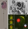
Size of this preview: 800 × 553 pixels. Other resolutions: 320 × 221 pixels | 640 × 442 pixels | 1,056 × 730 pixels.
Original file (1,056 × 730 pixels, file size: 863 KB, MIME type: image/png)
| This is a file from the Wikimedia Commons. Information from its description page there is shown below. Commons is a freely licensed media file repository. You can help. |
Summary
| Description |
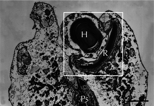
English: Electron micrograph of warnowiid oscelloid from Erythropsidinium, , highlighted in a white box. Positions of the hyalosome (H) and the retinal body (R) are indicated. A portion of the piston (Ps) is also visible.
Lightly edited to remove panel label from Figure 1B of Hayakawa S, Takaku Y, Hwang JS, Horiguchi T, Suga H, et al. (2015) Function and Evolutionary Origin of Unicellular Camera-Type Eye Structure. PLoS ONE 10(3): e0118415. doi: 10.1371/journal.pone.0118415 |
||
| Date | |||
| Source | Fig. 1B at http://journals.plos.org/plosone/article?id=10.1371/journal.pone.0118415 Function and Evolutionary Origin of Unicellular Camera-Type Eye Structure. PLoS ONE | ||
| Author | Shiho Hayakawa, Yasuharu Takaku, Jung Shan Hwang, Takeo Horiguchi, Hiroshi Suga, Walter Gehring, Kazuho Ikeo, Takashi Gojoboi | ||
| Permission (Reusing this file) |
|
||
| Other versions |
File history
Click on a date/time to view the file as it appeared at that time.
| Date/Time | Thumbnail | Dimensions | User | Comment | |
|---|---|---|---|---|---|
| current | 01:20, 24 July 2015 | | 1,056 × 730 (863 KB) | Opabinia regalis | == {{int:filedesc}} == {{Information |Description ={{en|1=Electron micrograph of warnowiid oscelloid from ''Erythropsidinium'', , highlighted in a white box. Positions of the hyalosome (H) and the retinal body (R) are indicated. A portion of the pis... |
File usage
The following pages on the English Wikipedia use this file (pages on other projects are not listed):
Global file usage
The following other wikis use this file:
- Usage on de.wikipedia.org
- Usage on pl.wikipedia.org
- Usage on ru.wikipedia.org