File:MultiPhotonExcitation-Fig4-doi10.1186slash1475-925X-5-36.JPEG

Original file (1,063 × 1,522 pixels, file size: 195 KB, MIME type: image/jpeg)

Summary

Description
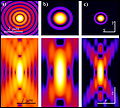
English: Original figure legend: MPE simplified optical pathways. In the MPE optical pathways the emission pinhole is removed since the only emitted light reaching the sensor is coming from the currently point scanned volume in the sample. No other fluorescence signal is produced elsewhere.
Date
Source

Multi-photon excitation microscopy. BioMedical Engineering OnLine, 2006, 5:36.
DOI:10.1186/1475-925X-5-36.

http://www.biomedical-engineering-online.com/content/5/1/36
Author Alberto Diaspro, Paolo Bianchini, Giuseppe Vicidomini, Mario Faretta, Paola Ramoino and Cesare Usai
Permission
(Reusing this file)
w:en:Creative Commons
attribution
This file is licensed under the Creative Commons Attribution 2.0 Generic license.
You are free:
  • to share – to copy, distribute and transmit the work
  • to remix – to adapt the work
Under the following conditions:
  • attribution – You must give appropriate credit, provide a link to the license, and indicate if changes were made. You may do so in any reasonable manner, but not in any way that suggests the licensor endorses you or your use.

All images uploaded from this article about multi-photon and two-photon-microscopy:

Captions

Add a one-line explanation of what this file represents

Items portrayed in this file

depicts

6 June 2006

File history

Click on a date/time to view the file as it appeared at that time.

Date/TimeThumbnailDimensionsUserComment
current18:08, 23 December 2008Thumbnail for version as of 18:08, 23 December 20081,063 × 1,522 (195 KB)Dietzel65== Beschreibung == {{Information |Description={{en|1=Original figure legend: ''MPE simplified optical pathways. In the MPE optical pathways the emission pinhole is removed since the only emitted light reaching the sensor is coming from the currently point

The following page uses this file:

Metadata