No higher resolution available.
Laminitis_radiograph_with_annotation.jpg (800 × 578 pixels, file size: 34 KB, MIME type: image/jpeg)
| This is a file from the Wikimedia Commons. Information from its description page there is shown below. Commons is a freely licensed media file repository. You can help. |
| Description |
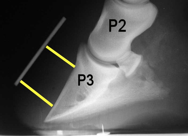
English: A typical x-ray or radiograph of a laminitic foot in a horse. The annotation P2 stands for the middle phalanx (pastern bone) and P3 denotes the distal phalanx (coffin bone). The white line marks the boundary of the outer hoof wall and the yellow lines show the distance between the top and bottom part of the coffin bone with the outer hoof wall. In this example the distal (bottom) part of the coffin bone is rotated away (greater distance) from the hoof wall, an indication of laminitis. |
||
| Date | |||
| Source | Radiograph created by Malcolm Morley , annotated by Froggerlaura | ||
| Author | Malcolm Morley | ||
| Permission (Reusing this file) |
|
File history
Click on a date/time to view the file as it appeared at that time.
| Date/Time | Thumbnail | Dimensions | User | Comment | |
|---|---|---|---|---|---|
| current | 04:00, 13 January 2012 | | 800 × 578 (34 KB) | Froggerlaura | {{Information |Description ={{en|1=A typical x-ray or radiograph of a laminitic foot in a horse. The annotation P2 stands for the middle phalanx (pastern bone) and P3 denotes the distal phalanx (coffin bone). The white line marks the boundary of the ou |
File usage
The following pages on the English Wikipedia use this file (pages on other projects are not listed):