Original file (1,310 × 647 pixels, file size: 89 KB, MIME type: image/jpeg)
Summary edit
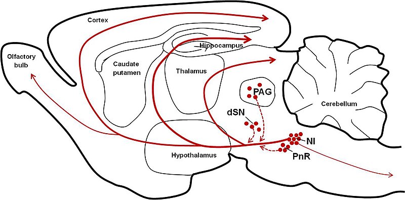
Parasagital schematic of the rodent brain displaying the major source of relaxin-3 neurons within the nucleus incertus (NI), while smaller populations are present within the pontine raphe (PnR), periaqueductal gray (PAG), and in a region dorsal to the substantia nigra (dSN). From here, relaxin-3 is trafficked along efferent projections throughout the brain. Image created by Craig Smith.
Licensing edit
| This work is licensed under the Creative Commons Attribution 3.0 License. |
 | This file is a candidate to be copied to Wikimedia Commons.
Any user may perform this transfer; refer to Wikipedia:Moving files to Commons for details. If this file has problems with attribution, copyright, or is otherwise ineligible for Commons, then remove this tag and DO NOT transfer it; repeat violators may be blocked from editing. Other Instructions
| ||
| |||
File history
Click on a date/time to view the file as it appeared at that time.
| Date/Time | Thumbnail | Dimensions | User | Comment | |
|---|---|---|---|---|---|
| current | 11:50, 11 April 2012 |  | 1,310 × 647 (89 KB) | Smittawiki (talk | contribs) | Parasagital schematic of the rodent brain displaying the major source of relaxin-3 neurons within the nucleus incertus (NI), while smaller populations are present within the pontine raphe (PnR), periaqueductal gray (PAG), and in a region dorsal to the ... |
You cannot overwrite this file.