File:Accessory and sesamoid bones of the foot - dorsoplantar projection.jpg

Original file(1,219 × 2,500 pixels, file size: 817 KB, MIME type: image/jpeg)

Summary

Description
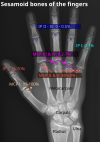
English: X-ray of the right foot by dorsoplantar projection, with areas indicating where the most common accessory and sesamoid bones would be, colored by their prevalences, and with representative shapes.
Date
Source

Own work
References
edit

Prevalences:

Os peroneum

  • Dorsoplantar view:
    • Brion Benninger, Jessica Kloenne (February 2011). "The Clinical Importance of the Os Peroneum: A Dissection of 156 Limbs Comparing the Incidence Rates in Cadavers versus Chronological Roentgenograms". The Foot and Ankle Online Journal 4 (2)., citing: (2008) (40th ed.), Elsevier ISBN: 9780702058455.
  • Lateral view:
    • (2013). "Prevalence of Accessory Ossicles and Sesamoid Bones in Hallux Valgus". Journal of the American Podiatric Medical Association 103 (3): 208–212. DOI:10.7547/1030208. ISSN 8750-7315.

Interphalangeal of great toe

  • Dorsoplantar and lateral view:
    • B. de Hartog; P.F. Doorn;P.C. Rijk (1). "Sesamoid bone interposition in the interphalangeal joint after dislocation of the hallux: A case report". The Foot and Ankle Online Journal 2 (7).

Os trigonum

  • Lateral view:
  • Not within the projection on dorsoplantar view.

Os tibiale externum or accessory navicular

Os intermetatarseum

Os vesalianum:

Os supranaviculare.

Os supratalare

Os talotibiale.

Os calcaneus secundarium

Metatarsophalangeal

Author Mikael Häggström
Other versions

edit


Versions and translations:

Foot, dorsoplantar Foot, lateral Ankle Wrist Fingers
Raster (.jpg)
Vector (.svg)
Translations
Source images

Licensing

I, the copyright holder of this work, hereby publish it under the following license:
Creative Commons CC-Zero This file is made available under the Creative Commons CC0 1.0 Universal Public Domain Dedication.
The person who associated a work with this deed has dedicated the work to the public domain by waiving all of their rights to the work worldwide under copyright law, including all related and neighboring rights, to the extent allowed by law. You can copy, modify, distribute and perform the work, even for commercial purposes, all without asking permission.

Captions

Add a one-line explanation of what this file represents

Items portrayed in this file

depicts

4 November 2017

image/jpeg

91b06857c6f4fb5e62ce948178a7026ae9b780e4

836,726 byte

2,500 pixel

1,219 pixel

File history

Click on a date/time to view the file as it appeared at that time.

Date/TimeThumbnailDimensionsUserComment
current18:48, 12 November 2017Thumbnail for version as of 18:48, 12 November 20171,219 × 2,500 (817 KB)Mikael HäggströmIn view
18:44, 12 November 2017Thumbnail for version as of 18:44, 12 November 20171,219 × 2,500 (817 KB)Mikael HäggströmAlso calcaneus
07:45, 9 November 2017Thumbnail for version as of 07:45, 9 November 20171,219 × 2,500 (816 KB)Mikael HäggströmNames of regular bones
12:19, 5 November 2017Thumbnail for version as of 12:19, 5 November 20171,219 × 2,500 (804 KB)Mikael HäggströmDark boxes to all
09:59, 5 November 2017Thumbnail for version as of 09:59, 5 November 20171,219 × 2,500 (802 KB)Mikael HäggströmCorrected: the supra-bones are behind on a dorsoplantar projection
18:40, 4 November 2017Thumbnail for version as of 18:40, 4 November 20171,224 × 2,500 (809 KB)Mikael HäggströmUser created page with UploadWizard
The following pages on the English Wikipedia use this file (pages on other projects are not listed):

Global file usage

The following other wikis use this file:

Metadata