This article was moved out to Anatomical terminology CFCF (talk) 21:15, 16 November 2013 (UTC)
This user page or section recently underwent a major revision or rewrite and may need further review. You can help Wikipedia by assisting in the revision. If this user page has not been edited in several days, please remove this template. This page was last edited by BD2412 (talk | contribs) 7 years ago. (Update timer) |

Anatomists and health care providers use anatomical terminology and medical terminology intermittently. These languages can be bewildering to the uninitiated, however the purpose of this language is not to confuse, but rather to increase precision and reduce medical errors. For example, is a scar “above the wrist” located on the forearm two or three inches away from the hand? Or is it at the base of the hand? Is it on the palm-side or back-side? By using precise anatomical terminology, ambiguity is eliminated. Anatomical terms derive from ancient Greek and Latin words, and because these languages are no longer used in everyday conversation, the meaning of their words does not change.[1]
Anatomical vocabulary edit
Anatomical terms are made up of roots, prefixes, and suffixes. The root of a term often refers to an organ, tissue, or condition, whereas the prefix or suffix often describes the root. For example, in the disorder hypertension, the prefix “hyper-” means “high” or “over,” and the root word “tension” refers to pressure, so the word “hypertension” refers to abnormally high blood pressure. The roots, prefixes and suffixes are often derived from Greek or Latin, and often quite dissimilar from their English-language variants.[1]
Latin names of structures such as musculus biceps brachii can be split up and refer to, musculus for muscle, biceps for "two-headed", brachii as in the brachial region of the arm. The first word tells us what we are speaking about, the second describes it, and the third points to location.[citation needed]
Use of the terms right and left edit
When anatomists refer to the right and left of the body, it is in reference to the right and left of the subject, not the right and left of the observer. When observing a body in the anatomical position, the left of the body is on the observer’s right, and vice versa.
Additionally the latin terms sinister for left, and dexter for right are used.[citation needed]
Relative location edit


Anatomical terminology is often chosen to highlight the relative location of body structures. For instance, an anatomist might describe one band of tissue as “inferior to” another or a physician might describe a tumor as “superficial to” a deeper body structure. Terms are used to define the relative location of body structures in a body that is positioned in the anatomical position, which is standing, feet apace, with palms forward and thumbs facing outwards. [1]
To further increase precision, anatomists standardize the way in which they view the body. Just as maps are normally oriented with north at the top, the standard body “map,” or anatomical position, is that of the body standing upright, with the feet at shoulder width and parallel, toes forward. The upper limbs are held out to each side, and the palms of the hands face forward. Using the standard anatomical position reduces confusion. It does not matter how the body being described is oriented, the terms are used as if it is in anatomical position. For example, a scar in the “anterior (front) carpal (wrist) region” would be present on the palm side of the wrist. The term “anterior” would be used even if the hand were palm down on a table.[1]
These standardized terms avoid confusion. Examples of terms include:
- Anterior and posterior, which describe structures at the front (anterior) and back (posterior) of the body. For example, the toes are anterior to the heel, and the popliteus is posterior to the patella.
- Superior and inferior, which describe a position above (superior) or below (inferior) another part of the body. For example, the orbits are superior to the oris, and the pelvis is inferior to the abdomen.
- Proximal and distal, which describe a position that is closer (proximal) or further (distal) from the trunk of the body. For example, the shoulder is proximal to the arm, and the foot is distal to the knee.
- Superficial and deep, which describe structures that are closer to (superficial) or further from (deep) the surface of the body. For example, the skin is superficial to the bones, and the brain is deep to the skull. Sometimes profound is used synonymously with deep.
- Medial and lateral, which describe a position that is closer to (medial) or further from (lateral) the midline of the body. For example, the nose is medial to the eyes, and the thumb is lateral to the other fingers.
All terms describe structures in a body that is in the anatomical position.
- Ventral and Dorsal, which describe structures derived from the front (ventral) and back (dorsal) of the embryo, before limb rotation.
- Superior (or cranial) describes a position above or higher than another part of the body proper.
- Inferior (or caudal) describes a position below or lower than another part of the body proper; near or toward the tail (in humans, the coccyx, or lowest part of the spinal column). [1]
Skull edit

Different terms are used when it comes to the skull in compliance with it's embryonic origin and its tilted position compared to in other animals.[citation needed]
- Rostral refers to the proximity to the front of the skull while
- Caudal refers to proximity to the base of the skull
Arm edit
When speaking of the arm different terminology is often used, so as to take account of the supination action the arm can perform. Therefor the terms ventral for anterior and dorsal for posterior are used preferentially. Aside from this additional terms are employed:[citation needed]
Planes edit

Anatomy is often described in planes, referring to a two-dimensional sections of the body. A section is a two-dimensional surface of a three-dimensional structure that has been cut. A plane is an imaginary two-dimensional surface that passes through the body. Three planes are commonly referred to in anatomy and medicine:
- The sagittal plane is the plane that divides the body or an organ vertically into right and left sides. If this vertical plane runs directly down the middle of the body, it is called the midsagittal or median plane. If it divides the body into unequal right and left sides, it is called a parasagittal plane or less commonly a longitudinal section.
- The frontal plane is the plane that divides the body or an organ into an anterior (front) portion and a posterior (rear) portion. The frontal plane is often referred to as a coronal plane, following Latin corona, which means "crown".
- The transverse plane is the plane that divides the body or organ horizontally into upper and lower portions. Transverse planes produce images referred to as cross sections.
Functional state edit
Anatomical terms may be used to describe the functional state of an organ:
- Anastamoses, the reconnection of two streams that previously branched out, such as blood vessels or leaf veins.[citation needed]
- Patent, meaning persistent after they would normally dissapear such as; Patent ductus arteriosus.
Regions edit
A body that is lying down is described as either prone or supine. Prone describes a face-down orientation, and supine describes a face up orientation. These terms are sometimes used in describing the position of the body during specific physical examinations or surgical procedures.[1]

The human body’s numerous regions have specific terms to help increase precision. Notice that the term “brachium” or “arm” is reserved for the “upper arm” and “antebrachium” or “forearm” is used rather than “lower arm.” Similarly, “femur” or “thigh” is correct, and “leg” or “crus” is reserved for the portion of the lower limb between the knee and the ankle.[1]
Body landmarks edit
Certain structures are used as landmarks on the body. Examples of such structures are the:
Body cavities edit

The body maintains its internal organization by means of membranes, sheaths, and other structures that separate compartments. The dorsal (posterior) cavity and the ventral (anterior) cavity are the largest body compartments. These cavities contain and protect delicate internal organs, and the ventral cavity allows for significant changes in the size and shape of the organs as they perform their functions. The lungs, heart, stomach, and intestines, for example, can expand and contract without distorting other tissues or disrupting the activity of nearby organs.[1]
The ventral cavity includes the thoracic and abdominopelvic cavities and their subdivisions. The dorsal cavity includes the cranial and spinal cavities. This illustration shows a lateral and anterior view of the body and highlights the body cavities with different colors.[1]
- The cranial cavity is a large, bean-shaped cavity filling most of the upper skull where the brain is located.
- The vertebral cavity is a very narrow, thread-like cavity running from the cranial cavity down the entire length of the spinal cord.
Together the cranial cavity and vertebral cavity can be referred to as the dorsal body cavity. In the posterior (dorsal) cavity, the cranial cavity houses the brain, and the spinal cavity (or vertebral cavity) encloses the spinal cord. Just as the brain and spinal cord make up a continuous, uninterrupted structure, the cranial and spinal cavities that house them are also continuous. The brain and spinal cord are protected by the bones of the skull and vertebral column and by cerebrospinal fluid, a colorless fluid produced by the brain, which cushions the brain and spinal cord within the posterior (dorsal) cavity.[1]
The thoracic cavity consists of three cavities that fill the interior area of the chest.
- The two pleural cavities are situated on both sides of the body, anterior to the spine and lateral to the breastbone.
- The superior mediastinum is a wedge-shaped cavity located between the superior regions of the two thoracic cavities.
- The pericardial cavity within the mediastinum is located at the center of the chest below the superior mediastinum. The pericardial cavity roughly outlines the shape of the heart.
The diaphragm divides the thoracic and the abdominal cavities. The abdominal cavity occupies the entire lower half of the trunk, anterior to the spine. Just under the abdominal cavity, anterior to the buttocks, is the pelvic cavity. The pelvic cavity is funnel shaped and is located inferior and anterior to the abdominal cavity. Together the abdominal and pelvic cavity can be referred to as the abdominopelvic cavity while the thoracic, abdominal, and pelvic cavities together can be referred to as the ventral body cavity.[1] Subdivisions of the Posterior (Dorsal) and Anterior (Ventral) Cavities The anterior (ventral) cavity has two main subdivisions: the thoracic cavity and the abdominopelvic cavity. The thoracic cavity is the more superior subdivision of the anterior cavity, and it is enclosed by the rib cage. The thoracic cavity contains the lungs and the heart, which is located in the mediastinum. The diaphragm forms the floor of the thoracic cavity and separates it from the more inferior abdominopelvic cavity. The abdominopelvic cavity is the largest cavity in the body. Although no membrane physically divides the abdominopelvic cavity, it can be useful to distinguish between the abdominal cavity, the division that houses the digestive organs, and the pelvic cavity, the division that houses the organs of reproduction.[1]
Abdomen edit

To promote clear communication, for instance about the location of a patient’s abdominal pain or a suspicious mass, health care providers typically divide up the cavity into either nine regions or four quadrants.[1]
Quadrants edit
The abdomen may be divided into four quadrants, more commonly used in medicine, subdivides the cavity with one horizontal and one vertical line that intersect at the patient’s umbilicus (navel).. The right upper quadrant (RUQ) includes the lower right ribs, right side of the liver, and right side of the transverse colon. The left upper quadrant (LUQ) includes the lower left ribs, stomach, spleen, and upper left area of the transverse colon. The right lower quadrant (RLQ) includes the right half of the small intestines, ascending colon, right pelvic bone and upper right area of the bladder. The left lower quadrant (LLQ) contains the left half of the small intestine and left pelvic bone. [1]
Regions edit
The more detailed regional approach subdivides the cavity with one horizontal line immediately inferior to the ribs and one immediately superior to the pelvis, and two vertical lines drawn as if dropped from the midpoint of each clavicle, resulting in nine regions. The upper right square is the right hypochondriac region and contains the base of the right ribs. The upper left square is the left hypochondriac region and contains the base of the left ribs. The epigastric region is the upper central square and contains the bottom edge of the liver as well as the upper areas of the stomach. The diaphragm curves like an upside down U over these three regions. The central right region is called the right lumbar region and contains the ascending colon and the right edge of the small intestines. The central square contains the transverse colon and the upper regions of the small intestines. The left lumbar region contains the left edge of the transverse colon and the left edge of the small intestine. The lower right square is the right iliac region and contains the right pelvic bones and the ascending colon. The lower left square is the left iliac region and contains the left pelvic bone and the lower left regions of the small intestine. The lower central square contains the bottom of the pubic bones, upper regions of the bladder and the lower region of the small intestine.[1]
Membranes edit

A serous membrane (also referred to as a serosa) is one of the thin membranes that cover the walls and organs in the thoracic and abdominopelvic cavities. The parietal layers of the membranes line the walls of the body cavity (pariet- refers to a cavity wall). The visceral layer of the membrane covers the organs (the viscera). Between the parietal and visceral layers is a very thin, fluid-filled serous space, or cavity.[1] Serous membrane lines the pericardial cavity and reflects back to cover the heart—much the same way that an underinflated balloon would form two layers surrounding a fist.Serous Membrane This diagram shows the pericardium on the left next to an analogy of a hand punching a balloon on the right. The pericardium is a two-layered sac that surrounds the entire heart except where the blood vessels emerge on the heart’s superior side. The pericardium has two layers because it folds over itself in the shape of the letter U. The inner layer that borders the heart is the visceral pericardium while the outer layer is the parietal pericardium. The space between the two layers is called the pericardial cavity. The heart sits in the cavity much like a fist punching into a balloon. The balloon surrounds the lower part of the fist with a two-layered sac, with the top of the balloon, where it contacts the fist, being analogous to the visceral pericardium. The bottom of the balloon, where it is tied off, is analogous to the parietal pericardium. The air within the balloon is analogous to the pericardial cavity.[1]
There are three serous cavities and their associated membranes. The pleura is the serous membrane that surrounds the lungs in the pleural cavity; the pericardium is the serous membrane that surrounds the heart in the pericardial cavity; and the peritoneum is the serous membrane that surrounds several organs in the abdominopelvic cavity. The serous fluid produced by the serous membranes reduces friction between the walls of the cavities and the internal organs when they move, such as when the lungs inflate or the heart beats. Both the parietal and visceral serosa secrete the thin, slippery serous fluid that prevents friction when an organ slides past the walls of a cavity. In the pleural cavities, pleural fluid prevents friction between the lungs and the walls of the cavity. In the pericardial sac, pericardial fluid prevents friction between the heart and the walls of the pericardial sac. And in the peritoneal cavity, peritoneal fluid prevents friction between abdominal and pelvic organs and the wall of the cavity. The serous membranes therefore provide additional protection to the viscera they enclose by reducing friction that could lead to inflammation of the organs.[1]
The serous membranes have two layers—parietal and visceral—surrounding a fluid filled space. Serous membranes cover the lungs (pleural serosa), heart (pericardial serosa), and some abdominopelvic organs (peritoneal serosa).[1]
Movement edit


Synovial joints allow the body a tremendous range of movements. Each movement at a synovial joint results from the contraction or relaxation of the muscles that are attached to the bones on either side of the articulation. The type of movement that can be produced at a synovial joint is determined by its structural type. While the ball-and-socket joint gives the greatest range of movement at an individual joint, in other regions of the body, several joints may work together to produce a particular movement. Overall, each type of synovial joint is necessary to provide the body with its great flexibility and mobility. There are many types of movement that can occur at synovial joints. Movement types are generally paired, with one being the opposite of the other. Body movements are always described in relation to the anatomical position of the body: upright stance, with upper limbs to the side of body and palms facing forward.[1]
General motion edit
| Adjusting angle between two parts |
Flexion – Bending movement that decreases the angle between two parts. Bending the elbow, or clenching a hand into a fist, are examples of flexion. When sitting down, the knees are flexed. Flexion of the hip or shoulder moves the limb forward (towards the anterior side of the body). Good examples of hip flexors are the rectus femoris, sartorius, iliacus, and psoas. Some knee flexors are the biceps femoris, semitendinosus, and semimembranosus, and some elbow flexors are the brachialis, biceps brachii, and brachioradialis. |
Extension – The opposite of flexion; a straightening movement that increases the angle between body parts. In a conventional handshake, the fingers are fully extended. When standing up, the knees are extended. Extension of the hip or shoulder moves the limb backward (towards the posterior side of the body). Elbow extensors include the triceps brachii and anconeus. The main muscles that extends the hip is the gluteus maximus. The muscles that extend the knee are the quadriceps group: the rectus femoris, vastus lateralis, vastus medialis, and vastus intermedius. |
| Adjusting relation to mid-line of body |
Abduction – A motion that pulls a structure or part away from the midline of the body (or, in the case of fingers and toes, spreading the digits apart, away from the centerline of the hand or foot). Abduction of the wrist is called radial deviation. Raising the arms laterally is an example of abduction. A good example for the arm is the deltoid. Some leg abductors are the gluteus medius and the gluteus minimus. |
Adduction – A motion that pulls a structure or part toward the midline of the body, or towards the midline of a limb. Dropping the arms to the sides, or bringing the knees together, are examples of adduction. In the case of the fingers or toes, adduction is closing the digits together. Adduction of the wrist is called ulnar deviation. The inner thigh houses some adductors, including the adductor brevis, adductor longus, adductor magnus, and pectineus. The latissimus dorsi is a good example for the humerus. |
| Rotating body parts |
Internal rotation (or medial rotation) of the shoulder or hip would point the toes or the flexed forearm inwards (towards the midline). The pectoralis major and subscapularis both medially rotate the humerus. The adductor longus and adductor brevis both medially rotate the thigh. |
External rotation (or lateral rotation) is the opposite of internal rotation. It would turn the toes or the flexed forearm outwards (away from the midline). The sartorius laterally rotates the femur. The infraspinatus and teres minor both laterally rotate the humerus. |
| Adjusting elevation |
Elevation – Movement in a superior direction. The upper muscle fibers of the trapezius aid in elevating the apex of the shoulder. |
Depression – Movement in an inferior direction, the opposite of elevation. Opposite to the upper fibers, the lower half of the trapezius aids in depressing the apex of the shoulder. |
Special motions of the hands and feet edit
| surfaces of the hands and feet | The palm (adj palmar) of the hand corresponds to the sole (adj plantar) of the foot. The adjective volar, used mainly in orthopaedics, is synonymous with palmar and plantar. As in palmar flexion and plantar flexion.[1] | The dorsum (back) of the hand corresponds to the dorsum (top) of the foot. As in dorsiflexionof the hand and foot.[1] |
| rotation of the forearm | Pronation – A rotation of the forearm that moves the palm from an anterior-facing position to a posterior-facing position, or palm facing down. This is not medial rotation as this must be performed when the arm is half flexed. (See also Pronator quadratus and Pronator teres muscle.) | Supination – The opposite of pronation, the rotation of the forearm so that the palm faces anteriorly, or palm facing up. The hand is supine (facing anteriorly) in the anatomical position. (See also Supinator muscle.) |
| bending of the entire foot | Dorsiflexion – Flexion of the entire foot superiorly, as if taking one's foot off an automobile pedal. | Plantarflexion – Extension of the entire foot inferiorly, as if pressing an automobile pedal. Occurs at ankle. |
| movement of the sole of the foot | Eversion – the movement of the sole of the foot away from the median plane. | Inversion – the movement of the sole towards the median plane (same as when an ankle is twisted). |
| compound movement of the foot | Pronation - a combination of abduction, eversion, and dorsiflexion. | Supination - a combination of adduction, inversion, and plantarflexion. |
Other special motions edit
| anterior/posterior movement – general | Protrusion – The anterior movement of an object. This term is often applied to the jaw. | Retrusion – The opposite of protrusion, moving a part posteriorly. |
| anterior/posterior movement – shoulders | Protraction – Anterior movement of the arms at the shoulders. | Retraction – Posterior movement of the arms at the shoulders. |
| motion within body (such as in blood vessels or the digestive system) | anterograde motion is in the normal direction of flow. (For example, passage of food from the mouth to the stomach.) | retrograde motion means reversed flow. (For example, gastric reflux.) |
Some additional motions without clear opposites are as follows:
- Rotation – A motion that occurs when a part turns on its axis. The head rotates on the neck, as in shaking the head 'no'.
- Circumduction – The circular (or, more precisely, conical) movement of a body part, such as a ball-and-socket joint or the eye. It consists of a combination of flexion, extension, adduction, and abduction. "Windmilling" the arms or rotating the hand from the wrist are examples of circumductive movement.
- Opposition – A motion involving a grasping of the thumb and fingers.
- Reposition – To release an object by spreading the fingers and thumb.
- Reciprocal motion of a joint – Alternating motion in opposing directions, such as the elbow alternating between flexion and extension.
- Occlusion - motion of the mandibula towards the maxilla making contact between the teeth.[2]
Types of joints edit
Movement is not limited to only synovial joints, although they allow for most freedom. Muscles also run over symphysis, which allow for movement in for example the vertebral column by compression of the intervertebral discs. Additionally synovial joints can be divided into different types, depending on their axis of movement;
| Type of joint | Example movements |
|---|---|
| Pivot Uniaxial joint | allows rotational movement Atlantoaxial joint (C1–C2 vertebrae articulation); proximal radioulnar joint |
| Hinge Uniaxial joint | allows flexion/extension movements Knee; elbow; ankle; interphalangeal joints of fingers and toes |
| Condyloid Biaxial joint | allows flexion/extension, abduction/adduction, and circumduction movements Metacarpophalangeal (knuckle) joints of fingers; radiocarpal joint of wrist; metatarsophalangeal joints for toes |
| Saddle Biaxial joint | allows flexion/extension, abduction/adduction, and circumduction movements First carpometacarpal joint of the thumb; sternoclavicular joint |
| Plane Multiaxial joint | allows inversion and eversion of foot, or flexion, extension, and lateral flexion of the vertebral column Intertarsal joints of foot; superior-inferior articular process articulations between vertebrae |
| Ball-and-socket Multiaxial joint | allows flexion/extension, abduction/adduction, circumduction, and medial/lateral rotation movements Shoulder and hip joints |
Muscle actions edit

Muscle action that moves the axial skeleton work over a joint with an origin and insertion of the muscle on respective side. The insertion is on the bone deemed to move towards the origin during muscle contraction. Muscles are often present that engage in several actions of the joint; able to perform for example both flexion and extension of the forearm as in the biceps and triceps respectively.[1] This is not only to be able to revert actions of muscles, but also brings on stability of the actions though muscle coactivation.[citation needed] The muscle performing an action is the agonist, while the muscle which contraction brings about an opposite action is the antagonist. For example a extension of the lower arm is performed by the triceps as the agonist and the biceps as the antagonist (which contraction will perform flexion over the same joint). Muscles that work together to perform the same action are called synergists.[1] In the above example synergists to the biceps can be the brachioradialis and the brachialis muscle.[citation needed]
Gross anatomy of muscles edit
This section needs additional citations for verification. (November 2013) |

The gross anatomy of a muscle is the most important indicator of its role in the body. One particularly important aspect of gross anatomy of muscles is pennation or lack thereof. In most muscles, all the fibers are oriented in the same direction, running in a line from the origin to the insertion. In pennate muscles, the individual fibers are oriented at an angle relative to the line of action, attaching to the origin and insertion tendons at each end. Because the contracting fibers are pulling at an angle to the overall action of the muscle, the change in length is smaller, but this same orientation allows for more fibers (thus more force) in a muscle of a given size. Pennate muscles are usually found where their length change is less important than maximum force, such as the rectus femoris.
Skeletal muscle is arranged in discrete muscles, an example of which is the biceps brachii. The tough, fibrous epimysium of skeletal muscle is both connected to and continuous with the tendons. In turn, the tendons connect to the periosteum layer surrounding the bones, permitting the transfer of force from the muscles to the skeleton. Together, these fibrous layers, along with tendons and ligaments, constitute the deep fascia of the body.
Systemic and regional approaches to anatomy edit
In the systematic approach to the study of anatomy, each system of the body, such as the digestive system, is studied as a thing in itself.[3] The regional approach observes all aspects of a particular region at one time.[3] For example, each muscle, organ, bone, nerve and any other structure in the abdomen is studied together, then the student continues in this fashion with each region. The regional approach better facilitates study with dissection, while the systematic approach is appreciated for its comprehensive look at each system throughout the body.
Additional images edit
-
 Older image of; Parts of the Human Body. Posterior and anterior view. From the 1933 Morris' Human Anatomy.
 -
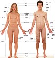 Human body features displayed on bodies on which body hair and male facial hair has been removed


See also edit
References edit
- ^ a b c d e f g h i j k l m n o p q r s t u v w x "Anatomy & Physiology". Openstax college at Connexions. Retrieved November 16, 2013.
- ^ "The Occlusion" (PDF). Retrieved November 15, 2013.
- ^ a b "Introduction page, "Anatomy of the Human Body". Henry Gray. 20th edition. 1918". Retrieved 19 March 2007.
This wikipedia entry incorporates text from the freely licenced Connexions [1] edition of Anatomy & Physiology[2] text-book by OpenStax College
-->