This article needs more reliable medical references for verification or relies too heavily on primary sources. (July 2018) |  |
A mucocele is a distension of a hollow organ or cavity because of mucus buildup.
By location edit
Oral edit
Oral mucocele is the most common benign lesion of the salivary glands generally conceded to be of traumatic origin. It is characterized by the pooling of mucus in a cavity due to the rupture of salivary ducts or acini. It can occur in the lower lip, palate, cheeks, tongue and the floor of the mouth.
Appendix edit
Appendiceal mucocele is found in 0.3 to 0.7% of the appendectomies.[1] It is characterized by the dilation of the organ lumen with mucus accumulation.[1] Appendix mucocele may come as a consequence of obstructive or inflammatory processes, cystadenomas or cystadenocarcinomas. Its main complication is pseudomyxoma peritonei.[1]
-
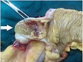 Gross pathology of mucocele of the appendix
 -
 Pie chart of histological types of mucocele of the appendix, with relative incidence.


Other edit
- Mucocele of the petrous apex
- Mucocele of the paranasal sinuses
- Gallbladder mucocele
Diagnosis edit
Superficial mucoceles can often be diagnosed by appearance and consistency alone. Sometimes, it is indicated to perform diagnostic imaging and/or needle biopsy.[citation needed]
On a CT scan, a mucocele is fairly homogenous, with an attenuation of about 10-18 Hounsfield units.[2]
See also edit
References edit
- ^ a b c B.B., Sunil Kumar; Jasuja, Pranav (2019). "Appendiceal mucocele—A rare case report". International Journal of Surgery Case Reports. 58: 21–25. doi:10.1016/j.ijscr.2019.04.008. ISSN 2210-2612. PMC 6468153. PMID 30999148.
- Creative Commons Attribution 4.0 International license - ^ page 152 in: Luca Saba and Jasjit S. Suri (2013). Multi-Detector CT Imaging: Principles, Head, Neck, and Vascular Systems. CRC Press. ISBN 9781439893845.