Chrysotile_SEM_photo.jpg (700 × 470 pixels, file size: 46 KB, MIME type: image/jpeg)
| This is a file from the Wikimedia Commons. Information from its description page there is shown below. Commons is a freely licensed media file repository. You can help. |
| Description |
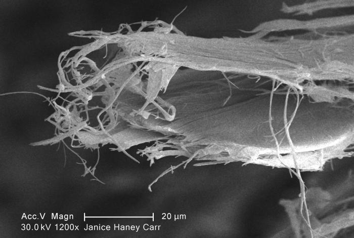
English: Under a high magnification of 1200X, this scanning electron micrograph (SEM) revealed some of the microcrystalline ultrastructure exhibited by a piece of raw chrysotile, or white asbestos, which had been excavated from the Lowell Asbestos Mine on Belvidere Mountain, Vermont.
Note the elongated crystalline structure, and how the fibrils are arranged in both bundles, and as singular serpentine units. See PHIL 11034 through PHIL 11066, for additional photomicrographic views of this material. The levels of asbestos in air that lead to lung disease depend on several factors. The most important of these are (1) how long you were exposed, (2) how long it has been since your exposure started, and (3) whether you smoked cigarettes. Cigarette smoking, and asbestos exposure increase your chances of getting lung cancer. Asbestos workers have increased chances of getting two principal types of cancer: cancer of the lung tissue itself and mesothelioma, a cancer of the thin membrane that surrounds the lung and other internal organs. These diseases do not develop immediately following exposure to asbestos, but appear only after a number of years. There is also some evidence from studies of workers that breathing asbestos can increase the chances of getting cancer in other locations (for example, the stomach, intestines, esophagus, pancreas, and kidneys), but this is less certain. Members of the public who are exposed to lower levels of asbestos may also have increased chances of getting cancer, but the risks are usually small and are difficult to measure directly. Lung cancer is usually fatal, while mesothelioma is almost always fatal, often within a few months of diagnosis. Some scientists believe that early identification and intervention of mesothelioma may increase survival. Copyright Restrictions: None - This image is in the public domain and thus free of any copyright restrictions. As a matter of courtesy we request that the content provider be credited and notified in any public or private usage of this image. |
|||
| Date | ||||
| Source | CDC PHIL image library, PHIL #11065 | |||
| Author | Janice Haney Carr | |||
| Permission (Reusing this file) |
|
File history
Click on a date/time to view the file as it appeared at that time.
| Date/Time | Thumbnail | Dimensions | User | Comment | |
|---|---|---|---|---|---|
| current | 02:13, 10 May 2010 | | 700 × 470 (46 KB) | Oaktree b | {{Information |Description={{en|1=Under a high magnification of 1200X, this scanning electron micrograph (SEM) revealed some of the microcrystalline ultrastructure exhibited by a piece of raw chrysotile, or white asbestos, which had been excavated from th |
File usage
Global file usage
The following other wikis use this file:
- Usage on af.wikipedia.org
- Usage on eu.wikipedia.org
- Usage on fr.wikipedia.org
- Usage on ru.wikipedia.org
- Usage on simple.wikipedia.org
- Usage on sl.wikipedia.org
- Usage on zh.wikipedia.org