Size of this preview: 632 × 600 pixels. Other resolutions: 253 × 240 pixels | 506 × 480 pixels | 809 × 768 pixels | 1,200 × 1,139 pixels.
Original file (1,200 × 1,139 pixels, file size: 165 KB, MIME type: image/jpeg)
| This is a file from the Wikimedia Commons. Information from its description page there is shown below. Commons is a freely licensed media file repository. You can help. |
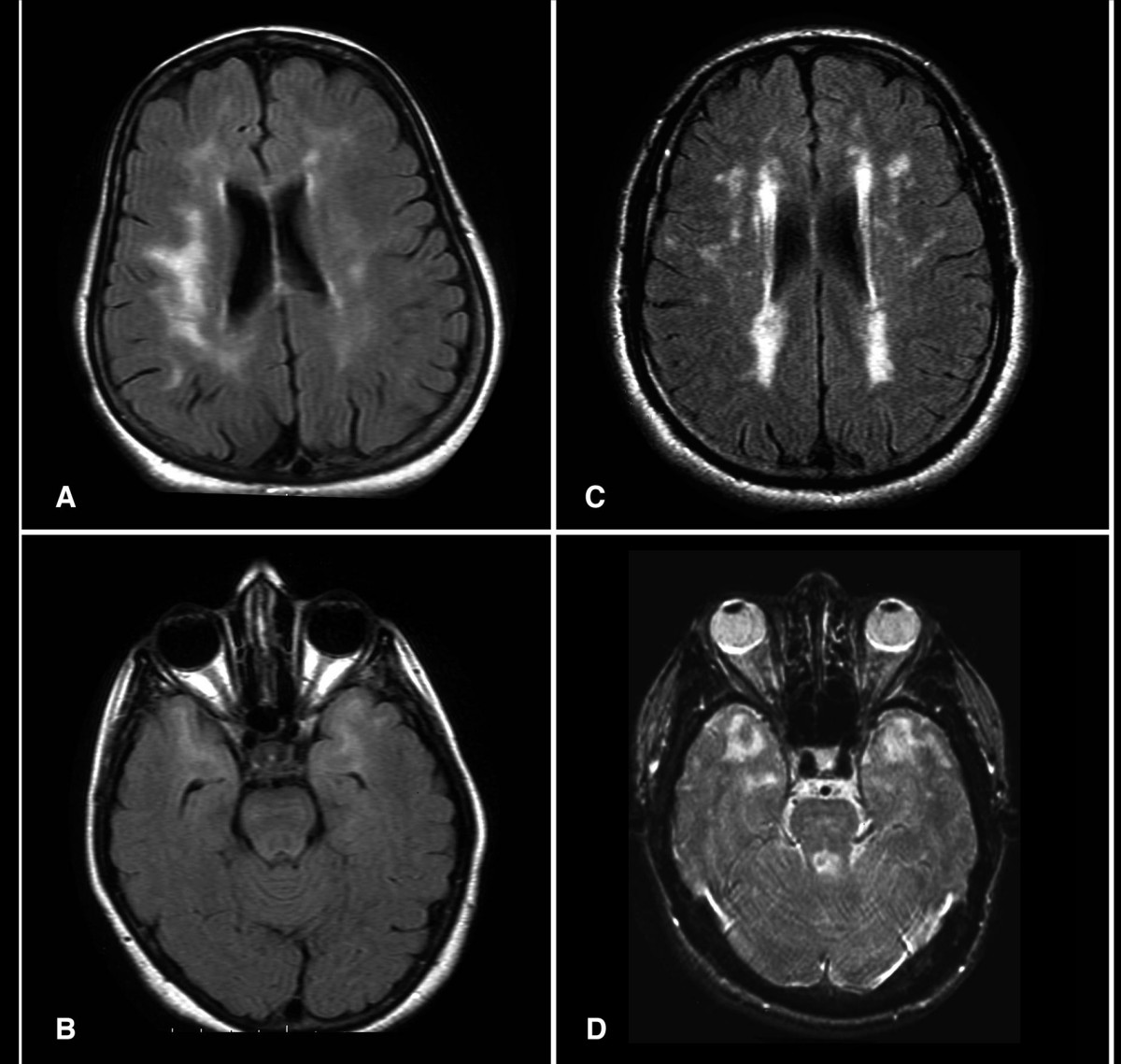
| Description | Axial FLAIR (a, b & c) and T2 weighted (d) Brain MRI from patients with CADASIL. The exams in 2a and 2b are from asymptomatic patients with depression. Note temporal lobe lesions even in asymptomatic patients (2b). In Figure 2a and d periventricular diffuse white matter ischemic lesion and multiple lacunar lesions in thalamus, pons and basal ganglia |
| Date | |
| Source | CADASIL in Arabs: clinical and genetic findings. BMC Medical Genetics 2007, 8:67doi:10.1186/1471-2350-8-67 |
| Author | Bohlega S, Al Shubili A, Edris A, Alreshaid A, Alkhairallah T, AlSous MW, Farah S, Abu-Amero KK. |
This file is licensed under the Creative Commons Attribution 2.0 Generic license.
- You are free:
- to share – to copy, distribute and transmit the work
- to remix – to adapt the work
- Under the following conditions:
- attribution – You must give appropriate credit, provide a link to the license, and indicate if changes were made. You may do so in any reasonable manner, but not in any way that suggests the licensor endorses you or your use.
File history
Click on a date/time to view the file as it appeared at that time.
| Date/Time | Thumbnail | Dimensions | User | Comment | |
|---|---|---|---|---|---|
| current | 12:18, 23 January 2008 | | 1,200 × 1,139 (165 KB) | Filip em | {{Information |Description=Axial FLAIR (a, b & c) and T2 weighted (d) Brain MRI from patients with CADASIL. The exams in 2a and 2b are from asymptomatic patients with depression. Note temporal lobe lesions even in asymptomatic patients (2b). In Figure 2a |
File usage
The following pages on the English Wikipedia use this file (pages on other projects are not listed):
Global file usage
The following other wikis use this file:
- Usage on ar.wikipedia.org
- Usage on bs.wikipedia.org
- Usage on ca.wikipedia.org
- Usage on da.wikipedia.org
- Bipolar affektiv sindslidelse
- Wikipedia:Dagens skandinaviske artikel/oktober 2010
- Wikipedia:Dagens skandinaviske artikel/Søndag/Uge 40, 2010
- Bruger:Hebbot/Sandkasse/WP:DSA/februar 2013
- Wikipedia:Dagens skandinaviske artikel/februar 2013
- Wikipedia:Dagens skandinaviske artikel/Svensk/Uge 7, 2013
- Wikipedia:Dagens skandinaviske artikel/Svensk/Uge 6, 2016
- Wikipedia:Dagens skandinaviske artikel/Søndag/Uge 6, 2017
- Usage on de.wikipedia.org
- Usage on es.wikipedia.org
- Usage on fa.wikipedia.org
- Usage on fi.wikipedia.org
- Usage on fr.wikipedia.org
- Usage on it.wikipedia.org
- Usage on ko.wikipedia.org
- Usage on la.wikipedia.org
- Usage on nl.wikipedia.org
- Usage on nn.wikipedia.org
- Wikipedia:Utvald svensk artikkel/2010
- Wikipedia:Utvald svensk artikkel/Veke 41, 2010
- Wikipedia:Utvald svensk artikkel/Veke 7, 2013
- Wikipedia:Utvald svensk artikkel/Veke 7, 2015
- Wikipedia:Utvald svensk artikkel/Veke 6, 2016
- Wikipedia:Utvald svensk artikkel/2013
- Wikipedia:Utvald svensk artikkel/2015
- Wikipedia:Utvald svensk artikkel/2016
- Usage on no.wikipedia.org
- Usage on pl.wikipedia.org
- Usage on ru.wikipedia.org
- Usage on sr.wikipedia.org
- Usage on sv.wikipedia.org
- Usage on www.wikidata.org