Barrel_cortex_pathways.jpg (400 × 457 pixels, file size: 82 KB, MIME type: image/jpeg)
| This is a file from the Wikimedia Commons. Information from its description page there is shown below. Commons is a freely licensed media file repository. You can help. |
| Description |
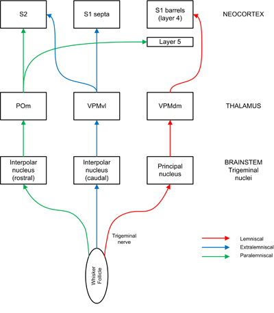
English: Sensory information flows in parallel pathways from whiskers to cortex. Sensory fibres from the whisker follicle run via the trigeminal nerve to brainstem nuclei, which project to nuclei in the contralateral thalamus, which in turn project to somatosensory cortex. The lemniscal pathway (red) runs via the principal nucleus and the dorsomedial section of the ventroposterior medial nucleus (VPMdm) to barrels in layer 4 of primary somatosensory cortex (S1). The extralemniscal pathway (blue) runs via the interpolar nucleus and the ventrolateral section of the VPM (VMPvl) to septa between barrels and to w:secondary somatosensory cortex (S2). The paralemniscal pathway (green) runs via the interpolar nucleus and the posterior nucleus (POm) to S2 and to barrel columns in S1 especially layer 5 (image by Peter Haslehurst, adapted from Diamond et al., 2008 and Fox, 2008). |
| Date | |
| Source | Own work |
| Author | Peterhaslehurst (talk) (Uploads) |
Sensory information flows in parallel pathways from whiskers to cortex. Sensory fibres from the whisker follicle run via the trigeminal nerve to brainstem nuclei, which project to nuclei in the contralateral thalamus, which in turn project to somatosensory cortex. The lemniscal pathway (red) runs via the principal nucleus and the dorsomedial section of the ventroposterior medial nucleus (VPMdm) to barrels in layer 4 of primary somatosensory cortex (S1). The extralemniscal pathway (blue) runs via the interpolar nucleus and the ventrolateral section of the VPM (VMPvl) to septa between barrels and to w:secondary somatosensory cortex (S2). The paralemniscal pathway (green) runs via the interpolar nucleus and the posterior nucleus (POm) to S2 and to barrel columns in S1 especially layer 5 (image by Peter Haslehurst, adapted from Diamond et al., 2008 and Fox, 2008).
| I, the copyright holder of this work, release this work into the public domain. This applies worldwide. In some countries this may not be legally possible; if so: I grant anyone the right to use this work for any purpose, without any conditions, unless such conditions are required by law. |

|
This media file is uncategorized.
Please help improve this media file by adding it to one or more categories, so it may be associated with related media files (how?), and so that it can be more easily found.
Please notify the uploader with {{subst:Please link images|File:Barrel cortex pathways.jpg}} ~~~~ |
File history
Click on a date/time to view the file as it appeared at that time.
| Date/Time | Thumbnail | Dimensions | User | Comment | |
|---|---|---|---|---|---|
| current | 16:25, 5 January 2009 | | 400 × 457 (82 KB) | Peterhaslehurst | Suppressed comment removed by FileImporter. |
| 16:21, 5 January 2009 |  | 3,525 × 4,026 (1.57 MB) | Peterhaslehurst | Sensory information flows in parallel pathways from whiskers to cortex. Sensory fibres from the whisker follicle run via the trigeminal nerve to brainstem nuclei, which project to nuclei in the contralateral thalamus, which in turn project to somatosensor |