The thoracodorsal nerve is a nerve present in humans and other animals, also known as the middle subscapular nerve or the long subscapular nerve. It supplies the latissimus dorsi muscle.[1][2]
| Thoracodorsal nerve | |
|---|---|
 Plan of brachial plexus. (Label for thoracodorsal nerve at bottom center.) | |
 Latissimus dorsi | |
| Details | |
| From | posterior cord (C6-C8) |
| Innervates | Latissimus dorsi muscle |
| Identifiers | |
| Latin | nervus thoracodorsalis |
| TA98 | A14.2.03.016 |
| TA2 | 6430 |
| FMA | 65290 |
| Anatomical terms of neuroanatomy | |
Structure edit
The thoracodorsal nerve arises from the brachial plexus. It derives its fibers from the sixth, seventh, and eighth cervical nerves. It is derived from their ventral rami, in spite of the fact that the latissimus dorsi is found in the back. The thoracodorsal nerve is a branch of the posterior cord of the brachial plexus,[1][3] and is made up of fibres from the posterior divisions of all three trunks of the brachial plexus.[4]
It follows the course of the subscapular artery, along the posterior wall of the axilla to the latissimus dorsi muscle,[1] in which it may be traced as far as the lower border of the muscle.
Function edit
The thoracodorsal nerve innervates the latissimus dorsi muscle on its deep surface.[1]
Clinical Significance edit
The latissimus dorsi is occasionally used for transplantation, and for augmentation of systole in cardiac failure. In these cases, the nerve supply is preserved, and transplanted with the muscle (for example, with facial reanimation).[5]
Posterior cord lesions can result in the loss of adduction of the shoulder joint, as innervation to latissimus dorsi is lost.[3]
Additional images edit
This gallery of anatomic features needs cleanup to abide by the medical manual of style. |
-
 Brachial plexus
 -
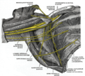 The right brachial plexus (infraclavicular portion) in the axillary fossa; viewed from below and in front.
 -
 Brachial plexus with courses of spinal nerves shown

References edit
![]() This article incorporates text in the public domain from page 934 of the 20th edition of Gray's Anatomy (1918)
This article incorporates text in the public domain from page 934 of the 20th edition of Gray's Anatomy (1918)
- ^ a b c d Nicole Bentley, J.; Yang, Lynda J. -S. (2015-01-01), Tubbs, R. Shane; Rizk, Elias; Shoja, Mohammadali M.; Loukas, Marios (eds.), "Chapter 42 - Anatomy of the Posterior Cord and Its Branches", Nerves and Nerve Injuries, San Diego: Academic Press, pp. 563–574, doi:10.1016/b978-0-12-410390-0.00012-3, ISBN 978-0-12-410390-0, retrieved 2020-11-01
- ^ Katirji, Bashar (2007-01-01), Katirji, Bashar (ed.), "Case 11", Electromyography in Clinical Practice (Second Edition), Philadelphia: Mosby, pp. 175–187, doi:10.1016/b978-0-323-02899-8.50020-3, ISBN 978-0-323-02899-8, retrieved 2020-11-01
- ^ a b Preston, David C.; Shapiro, Barbara E. (2013-01-01), Preston, David C.; Shapiro, Barbara E. (eds.), "30 - Brachial Plexopathy", Electromyography and Neuromuscular Disorders (Third Edition), London: W.B. Saunders, pp. 468–486, doi:10.1016/b978-1-4557-2672-1.00030-1, ISBN 978-1-4557-2672-1, retrieved 2020-11-01
- ^ Bertorini, Tulio E. (2008-01-01), Bertorini, Tulio E. (ed.), "1 - Neuromuscular Anatomy and Function", Neuromuscular Case Studies, Philadelphia: Butterworth-Heinemann, pp. 1–25, doi:10.1016/b978-0-7506-7332-7.50005-2, ISBN 978-0-7506-7332-7, retrieved 2020-11-01
- ^ Rea, Paul (2015-01-01), Rea, Paul (ed.), "Chapter 2 - Upper Limb Nerve Supply", Essential Clinically Applied Anatomy of the Peripheral Nervous System in the Limbs, Academic Press, pp. 41–100, doi:10.1016/b978-0-12-803062-2.00002-4, ISBN 978-0-12-803062-2, retrieved 2020-11-01
External links edit
- Dissection Video of Superficial Back showing the Thoracodorsal Nerve
- Anatomy figure: 05:03-10 at Human Anatomy Online, SUNY Downstate Medical Center - "The major subdivisions and terminal nerves of the brachial plexus."
- Thoracodorsal Nerve - BlueLink Anatomy - University of Michigan Medical School