The external anal sphincter (or sphincter ani externus) is an oval tube of skeletal muscle fibers.[1] Distally, it is adherent to the skin surrounding the margin of the anus.[2] It exhibits a resting state of tonical contraction.[1]
| External anal sphincter | |
|---|---|
 | |
 Coronal section through the anal canal. B. Cavity of urinary bladder V.D. Ductus deferens. S.V. Seminal vesicle. R. Second part of rectum. A.C. Anal canal. L.A. Levator ani. I.S. Sphincter ani internus. E.S. Sphincter ani externus. | |
| Details | |
| Nerve | Branch from the fourth sacral and contributions from the inferior hemorrhoidal branch of the pudendal nerve |
| Actions | Keep the anal canal and orifice closed |
| Identifiers | |
| Latin | sphincter ani externus |
| TA98 | A04.5.04.012 |
| TA2 | 2426 |
| FMA | 21930 |
| Anatomical terms of muscle | |
Anatomy edit
The external anal sphincter is far more substantial than the internal anal sphincter. The proximal portion of external anal sphincter overlaps the internal anal sphincter (which terminates distally a little distance proximal to the anal orifice) superficially; where the two overlap, they are separated by the intervening conjoint longitudinal muscle.[1]
Structure edit
Historically, the sphincter was described as consisting of three parts (deep, superficial, and subcontinuous). This is not supported by current anatomical knowledge. Some sources still describe it in two layers, deep (or proximal) and superficial (or distal or subcutaneous).[1]
Some of the muscles fibres decussate at the anterior midline and posterior midline, so forming an anterior commissure and posterior commissure.[1]
Attachments edit
This section needs expansion. You can help by adding to it. (July 2023) |
The muscle attaches anteriorly onto the perineal body, and posteriorly onto the anococcygeal ligament.[1]
Innervation edit
The sphincter receives innervation from the bilaterally paired inferior anal nerve (each a branch of the pudendal nerve which is derived from ventral rami of S2-S4). It may also receive additional motor innervation from the nerve to levator ani.[1]
Histology edit
The sphincter consists mostly of slow twitch fibers that allow extended continuous contraction.[1]
Gallery edit
-
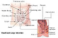 Intestines
 -
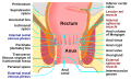 Anatomy of the human anus.
 -
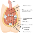 Muscles of male perineum.
 -
 Muscles of the female perineum.
 -
 Sagittal (vertical) section of bladder, penis, and urethra.


See also edit
References edit
- ^ a b c d e f g h Standring, Susan (1201). Gray's Anatomy: The Anatomical Basis of Clinical Practice (42th ed.). New York. p. 683. ISBN 978-0-7020-7707-4. OCLC 1201341621.
- ^ Gray, Henry (1918). Gray's Anatomy (20th ed.). pp. 424–425.
![]() This article incorporates text in the public domain from page 425 of the 20th edition of Gray's Anatomy (1918)
This article incorporates text in the public domain from page 425 of the 20th edition of Gray's Anatomy (1918)
External links edit
- Anatomy photo:42:13-0100 at the SUNY Downstate Medical Center - "The Male Perineum and the Penis: The External Anal Sphincter"
- perineum at The Anatomy Lesson by Wesley Norman (Georgetown University) (analtriangle3)
- pelvis at The Anatomy Lesson by Wesley Norman (Georgetown University) (rectum)