There are three cuneiform ("wedge-shaped") bones in the human foot:
- the first or medial cuneiform
- the second or intermediate cuneiform, also known as the middle cuneiform
- the third or lateral cuneiform
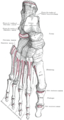
| Cuneiform bones; Cuneiform | |
|---|---|
 Red=medial; yellow=intermediate; green=lateral | |
 Cuneiform bones of the left foot | |
| Details | |
| Identifiers | |
| Latin | os cuneiformis pl. ossa cuneiformia |
| FMA | 24517 |
| Anatomical terms of bone | |
They are located between the navicular bone and the first, second and third metatarsal bones and are medial to the cuboid bone.[1]
Structure edit
There are three cuneiform bones:
- The medial cuneiform (also known as first cuneiform) is the largest of the cuneiforms. It is situated at the medial side of the foot, anterior to the navicular bone and posterior to the base of the first metatarsal. Lateral to it is the intermediate cuneiform. It articulates with four bones: the navicular, second cuneiform, and first and second metatarsals. The tibialis anterior and fibularis longus muscle inserts at the medial cuneiform bone.[2]
- The intermediate cuneiform (second cuneiform or middle cuneiform) is shaped like a wedge, the thin end pointing downwards. The intermediate cuneiform is situated between the other two cuneiform bones (the medial and lateral cuneiforms), and articulates with the navicular posteriorly, the second metatarsal anteriorly and with the other cuneiforms on either side.
- The lateral cuneiform (also known as third cuneiform or external cuneiform) intermediate in size between the other two cuneiform bones, is also wedge-shaped, the base being uppermost. It occupies the center of the front row of the tarsal bones, between the intermediate cuneiform medially, the cuboid laterally, the navicular posteriorly and the third metatarsal in front. The tibialis posterior inserts at the lateral cuneiform, while the flexor hallucis brevis originates from it.[2]
Muscle attachments edit
| Muscle | Direction | Attachment[2] |
| Tibialis anterior | Insertion | Medial cuneiform |
| Fibularis longus | Insertion | Medial cuneiform |
| Tibialis posterior | Insertion | Medial cuneiform |
| Flexor hallucis brevis | Origin | Lateral cuneiform |
Injuries edit
- Lisfranc fracture – in which one or all of the metatarsals are displaced from the tarsus[3]
- Cuneiform fracture - Due to the ligamentous support of the midfoot, isolated cuneiform fractures are rare [4]
Additional images edit
-
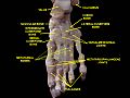
 Bones of the right foot. Dorsal surface.
 -
 Bones of the right foot. Plantar Surface.
 -
 Skeleton of foot. Medial aspect.
 -
 Skeleton of foot. Lateral aspect.
 -
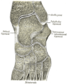 Oblique section of left intertarsal and tarsometatarsal articulations, showing the synovial cavities.
 -
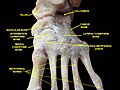
 Bones of foot
 -
 Cuneiform. Superior view.
 -
 Cuneiform. Superior view.


Other animals edit
This section needs expansion. You can help by adding to it. (April 2015) |
See also edit
- Cuneiform, for writing by pressing a wedge-shaped reed into wet clay.
References edit
Wikimedia Commons has media related to Cuneiform bones.
- ^ Bojsen-Møller, Finn; Simonsen, Erik B.; Tranum-Jensen, Jørgen (2001). Bevægeapparatets anatomi [Anatomy of the Locomotive Apparatus] (in Danish) (12th ed.). p. 245. ISBN 978-87-628-0307-7.
- ^ a b c Bojsen-Møller, Finn; Simonsen, Erik B.; Tranum-Jensen, Jørgen (2001). Bevægeapparatets anatomi [Anatomy of the Locomotive Apparatus] (in Danish) (12th ed.). pp. 364–367. ISBN 978-87-628-0307-7.
- ^ TheFreeDictionary > Lisfranc's fracture Citing: Mosby's Medical Dictionary, 8th edition. 2009
- ^ Mabry LM, Patti TN, Ross MD, Bleakley CM, Gisselman AS (July 2021). "Isolated Medial Cuneiform Fractures: A Systematic Search and Qualitative Analysis of Case Studies". J Am Podiatr Med Assoc. 111 (4): 1–9. doi:10.7547/20-047. PMID 34478529. S2CID 225705519.
{{cite journal}}: CS1 maint: multiple names: authors list (link)